
Special Article - Prosthodontics
Austin J Dent. 2018; 5(6): 1121.
Comparing the Incidence of Chipping in Tooth- Supported and Implant-Supported Ceramically Veneered Restorations
Brandt S¹*, Lochmann CH², Lauer H-CH¹, Brandt J¹ and Winter A³
¹Department of Prosthodontics, School of Dentistry Goethe University Frankfurt am Main, Germany
²Dentist in private practice, Bruchköbel, Germany
³Department of Prosthodontics, School of Dentistry Julius Maximilians University Würzburg, Germany
*Corresponding author: Brandt S, Department of Prosthodontics, School of Dentistry Goethe University Frankfurt am Main, Germany
Received: October 29, 2018; Accepted: November 26, 2018; Published: December 03, 2018
Abstract
Purpose: Chipping is very common in dentistry, with up to 40% of ceramically veneered restorations being affected. The purpose of the study was to examine possible differences in chipping rates between tooth-supported and implant-supported restorations in the same environments.
Materials and Methods: A total of 563 restorations in 148 patients were clinically re-examined. The study included only patients with a tooth-supported and an implant-supported ceramically veneered restoration placed within a limited time of each other.
Results: Both the tooth-supported and the implant-supported restorations exhibited chipping. However, there was no significant difference in chipping rates between tooth-supported and implant-supported ceramically veneered restorations. Also the region of the crown does not depend significant to the chipping.
Conclusions: Factors such as appropriate processing of the ceramic materials, polishing of finished or adjusted surfaces prior to delivery, and providing night guards for e.g. bruxism patients have a much greater impact on the incidence of chipping than whether the crown was placed on a natural tooth or an implant.
Keywords: Tooth and implant abutments; Chipping; Veneered restoration
Abbreviations
CAD/CAM: Computer Aided Design/Computer Aided Manufacturing; e.g.: for example; Fig: Figure; MPa: Megapascal; N: Newton
Introduction
The use of ceramics in dentistry was firstly mentioned 1733 in Fauchard’s book Französischer Zahn-Arzt , oder Traktat von den Zähnen “French Dentist, or Treatise of the Teeth”. He describes a procedure for firing ceramic veneers onto fixed partial dentures made of gold. However, his technique did not gain much ground due to the insufficient bond of the two materials [1].
Thanks to manifold improvements in chemical compositions and processing techniques, ceramics have now become established as an important material in restorative dentistry, combining excellent esthetics with high biocompatibility and a low affinity to plaque [2].
Dental ceramics can be subdivided into two main categories (oxide ceramics and silicate ceramics) with further subdivisions. Due to their stability and favorable shade, oxide ceramics are perfectly suited as framework materials, either fully or partially veneered or in fully contoured crowns [3]. Modern oxide ceramics are characterized by phase-transformation consolidation, a process in which the ceramic is stabilized in its tetragonal phase by oxide additives [4].
If cracks occur within the ceramics, the associated stress-induced release of energy causes a transformation from the tetragonal to the monoclinic phase [5]. The volume increase of 3%-5% halts the cracking [6]. Together with the high flexural strength (up to 1,200MPa) and cracking resistance (9-10 MPa m-1), this property makes oxide ceramics resistant to damage to a certain extent [7].
Silicate ceramics, which come in different translucencies and opacities, have a clear advantage over oxide ceramics in the esthetic zone [8]. Their generally low bending strength (approx. 100MPa) can be increased to up to 450MPa by adding lithium or a similar material. This added stability is accompanied by an increase in opacity, at the expense of esthetics [9].
All ceramic materials, despite their fundamental differences, must be able to resist the functional (and parafunctional) loads of the masticatory system. The maximum physiological load in the molar region, as measured in the normal dentition, is around 250N, according to Fernandes, et al. [10]. For implant-supported restoration, Morneburg, et al. reported values of 264-284 N [11]. These masticatory forces that impact the ceramics manifest themselves as compressive and tensile stress as well as shear forces. Compressive stress is much more readily tolerated by ceramic materials than tensile stress. When the forces applied exceed the elastic limits of the framework material, fracture becomes inevitable. This brittle fracture behavior, often mentioned in the literature, is to be attributed to the plastic deformation of ceramics, which is not present in metals [12].
Ceramic fractures can be cohesive or adhesive in nature. A cohesion fracture (Figure 1) is defined as a fracture within the veneering ceramics, without an imminent framework exposure. An adhesion fracture is defined as a fracture at the veneer/framework interface (Figure 2), resulting in parts of the framework being exposed.
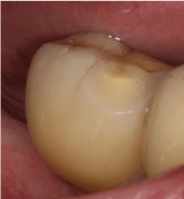
Figure 1: Cohesion fracture.
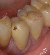
Figure 2: Adhesion fracture.
These fractures, jointly referred to as chipping, are a common issue in implant prosthetics according to Ozcan [13], which has been confirmed by several studies on this subject [14-16] and reports of chipping rates of up to 40% [17].
Veneer fractures in the posterior jaw are often accompanied by a loss in chewing efficiency, whereas chipping in the anterior jaw is mostly a purely esthetic problem [18]. Fractures of prosthetic frameworks are rare [19].
Previous studies have generally examined only tooth-supported or only implant-supported crowns. Strikingly, implant-supported restorations appear to be more prone to chipping than toothsupported ones. Kinsel and Lin [20] reported a chipping rate of 19.5% in implant-supported fixed partial dentures over an observation period of more than five years, whereas Eschbach, et al. [21] reported a fracture rate of 6% for tooth-supported fixed partial dentures over a similar period. Furthermore, the age and type of restoration [20,22,23], the type of connection (cemented or screw-retained) [24] and the framework material all seem to have an impact on a restoration’s proneness to fracture [15]. Numerous possible reasons for this have been mentioned in the literature [25]:
- Inadequate tooth preparation [26]
- Unsuitable framework materials [27] and shapes
- Mismatches in thermal expansion coefficients
- Firing inadequacies
- Insufficient wall thickness of the veneer [28]
- Possible problems within the ceramic material (pores, bubbles, etc.)
- Post-processing of the restoration (adjustments, finishing, polishing) [23]
None of the several extant publications on chipping directly compares the chipping behavior of tooth-supported and implantsupported ceramic restorations within one patient. The Department of Prosthetic Dentistry at the University of Frankfurt, Germany therefore conducted a study that facilitated a direct intraindividual comparison.
Materials and Methods
Approval for the study was sought and obtained from the responsible ethical review committee. Accounting data were consulted to identify potential study participants (searching for evidence of delivery of tooth- or implant-supported restorations). One aspect to be considered was that the delivery dates of the restorations examined should not be further apart than nine months to ensure equal environmental conditions for the restorations in the oral cavity (Figure 3).
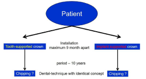
Figure 3: Identical concept and design.
The ensuing review of the medical records yielded was 189 eligible subjects. Of these, 148 patients agreed to participate after having been contacting them by phone or in writing. The patients included had received several ceramically veneered all-ceramic or metal-ceramic crowns, both implant-supported and tooth-supported. Thus, 563 crowns were examined. Appointments were made for detailed dental examinations. The subjects were -informed of the impending procedures. Their restorations were checked for possible chipping. The clinical findings were reported on a specific form consisting of four sections:
1. Patient data (oral examination, occlusion, abrasion)
2. Tooth-related data (region, age and type of restoration, surface processing, occlusion, chipping, gingivitis, mobility, morphology, antagonists, type of connection)
3. Implant-related data (region, age and type of restoration, surface processing, occlusion, chipping, gingivitis, mobility, morphology, antagonists, type of connection)
4. Laboratory data
Instances of chipping reported under sections 2 and 3 were additionally documented photographically.
The data on the reporting forms were entered into an Access database (Microsoft; Redmond, WA, USA) and tagged (1 for true and 2 for false), then analyzed using the SPSS Statistics and BiAS 9.14 software packages (IBM, Armonk, NY, USA).
A Kaplan Meier Analysis illustrates the survival time (until the first chipping occurred) and rate of the crowns. The Log-Rang- Test was used to check a statistical significance (p=0.05). Thoroughly literature research lead to the assumption of a defect probability of 10% for tooth-supported crowns and 25% for implant-supported crowns. Based on these data, a minimum number of cases for toothsupported (107) and implant-supported (107) crowns was calculated using the chi-square test.
Results
A total of 563 restorations in 148 patients were re-examined, of which 277 were tooth-supported and 286 implant-supported.
Chipping was detected in 19 of 277 tooth-supported crowns (6.9%) and in 22 of 286 implant-supported crowns (7.8%). There’s no statistical significance between the chipping of tooth and implant borne crowns (Log-Rang-Test p=0.884) The Kaplan Meier Analysis shows a mean survival time of implant borne crowns in general of 122,2 ± 2.1 months, of tooth borne crowns 118.2 ± 3,1 (Figure 4).
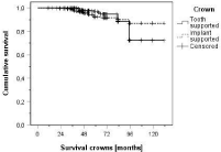
Figure 4: Kaplan-Meier analysis.
There’s also no significant difference of chipping neither between upper and lower jaw (p=0.882, Log-Rang-Test) nor between the regions (p=0.685) in general. Furthermore, there’s a lack of statistical significance of the regions between tooth - and implant supported crowns (tooth: p=0.940, implant: p=0.707).
Table 1 shows the mean survival time of the Kaplan Meier Analysis depending on the region. There occurred no chipping in the front tooth region.
![]()
    Region
    Upper jaw
    Lower jaw


    Tooth-premolar region
    125.1 ± 3.3
    92.4 ± 1.3


    Tooth-molar region
    102.4 ± 3
    99.5 ± 2.9


    Implant-premolar region
    120.8 ± 4.8
    94.4 ± 1.6


    Implant-molar region
    111.9 ± 3.9
    110.6 ±2.6
Table 1: Kaplan-Meier analysis.
In general, the older the restoration, the higher is the chipping rate. Thus, the Kaplan Meier Survival rate of the tooth borne crowns shows a 100% survival rate up to 41 months. The first chipping occurred after 42 months. Thus, the survival rate after 48 months is 97,7%, 97% after 60 months, 94,8% after 72 months, 88,5% after 84 months and 72,4% up to 131 months.
The implant borne crowns show a 100% survival rate up to 28 months. Afterwards values appear of 98,5% (36 months), 95,6% (48 months), 92,4% (60 months), 90,1% (84 months) and 86,9% after 131 months.
In 126 of 148 subjects there occurred no chipping. At 9 subjects there was at least one instance of chipping. In 7 cases occurred two chippings, 4 out of those 7 showed at least one instance of chipping each in a tooth-supported and an implant-supported restoration. The remaining number of patients suffered more than two chippings.
Discussion
The highly restrictive subject selection allowed a direct comparison of tooth-supported and implant-supported restorations. It ensured that the restorations examined were comparable in terms of the materials used, the manufacturer (dental technician) involved, and the time of exposure to the oral cavity.
Generally it should be mentioned, however, that the chipping may have been caused by earlier premature contacts no longer present at the time of examination. This problem in dental examination could be one of the reasons why prior studies rarely investigated the type of dynamic occlusion and the consequences of present abrasions in relation to the probability of chipping.
The fact that the region does not significant depend on the chipping of tooth- and implant- supported restorations can be explained by the manufacturing process of the crowns.
In general for crowning, the tooth has to be prepared. The aim of the preparation is an undersized form of the tooth and also the later framework has to be formed similar to it. This ensures an adequate support for the veneering material and can explain the low chipping rate of the tooth supported crowns.
In addition, at this study individual abutments were used for the implant supported crowns. They were designed and afterwards manufactured by the CAD/CAM technique. The design of these abutments imitates the prepared stump, similar to the undersized anatomic form of the original tooth. In this way also the framework can be produced full anatomically and can support the veneering material adequate.
Numerous authors reported a, sometimes considerable, increase in the incidence of chipping with restoration age [20,22,23]. This is only partly corroborated by the results of the present study. The tooth supported crowns show the first chipping after 42 months, In the following years, the number of chippings increased. Thus, 35.3% exhibited chipping over a time span of up to five years, whereas 64.7% of the restorations showed a chipping in the following three years. At implant-supported crowns, the first chipping occurred earlier after 29 months and also more crowns chipped in the time span of up to five years (55.8%). The different time of chipping might possibly find its explanation when taking a closer look at the framework materials of all crowns with chipping. In the case of tooth-supported restorations, all crowns affected by chipping had a metal framework, whereas in the case of implant-supported restorations, only 15 out of the 22 chipped restorations were metal-ceramic crowns.
It must therefore be assumed that fatigue of the adhesive bond between the metal framework and ceramic veneer develops as the restorations age. In addition, undersized metal frameworks (caused, for example, by a wish to save on the high cost of precious metals) can increase the risk of chipping. A supportive framework design and diligent firing (considering all requisite parameters) are necessary to reduce as far as possible the higher risk of chipping with increasing age.
In the course of this, the Kaplan Meier Analysis shows a 5-yearsurvival rate of 92.4% of implant supported crowns and 97% of tooth supported crowns. This corresponds to findings of Fradeani, et al. [29] and Gehrt, et al. [30]. They describe similar survival rates of 97.1% and 96.8% of tooth borne single crowns.
The survival rate for implant borne single crowns only partly corresponds with existing studies. Here, a meta-analysis by Pjetursson, et al. shows a higher survival rate of 94.5% after 5 years [31]. But Palmer, et al. support the findings of this study by describing a survival rate of 92.7% [32].
Looking at the total incidence of chippings in tooth-supported (and implant-supported crowns, the overall chipping frequency is in the same range as in prior studies. The implicit baseline hypothesisthat implant-supported crowns would exhibit significantly more chipping than tooth-supported ones-cannot be confirmed, however. A study by Sorrentino, et al. [33], investigated 128 tooth-supported and 81 implant-supported restorations six years retrospectively. Despite differences in selection criteria and a purely group-based analysis, no considerable difference was found in the chipping frequencies of tooth-supported and implant-supported restorations. Other studies relate to the chipping frequency of either toothsupported or implant-supported crowns, not both, and therefore do not allow a valid comparison [15,20,21,24].
In the case of patients with an increased probability of chipping (such as bruxism patients) [20], the use of alternative materialslithium disilicate or fully contoured zirconia-should be considered. If ceramically veneered crowns are nevertheless chosen in the end, obligatory preventive steps should be taken, such as a particularly meticulous occlusal adjustment, the polishing of occlusal-adjustment areas (or a final firing), or providing a nightguard [34].
Conclusion
The present study has falsified the hypothesis that ceramic chipping is more probable in implant-supported crowns, as often suggested by prior studies. Factors such as an anatomic framework and abutment design, appropriate post-processing of ceramic surfaces, or sufficient tooth preparation have a much greater impact on chipping and its avoidance than the abutment type of the restoration.
The positive effect of a final firing before delivery is not to be underestimated. The contrary results regarding the correlation between the age of a restoration and chipping probability show this previously problematic factor can be significantly defused by appropriate techniques and materials.
It should be possible to further decrease the chipping rates by considering the use of alternative materials, such as lithium disilicate or fully contoured zirconia.
References
- Tholey MJ, Thiel N. The burning of dental veneering ceramic. Quintessence dental technology. 2009: 1018-1029.
- Fischer H, Karaca F, Marx R. Detection of microscopic cracks in dental ceramic materials by fluorescent penetrant method. J Biomed Mater Res. 2002; 61: 153-158.
- Ramos GF, Monteiro EB, Bottino MA, Zhang Y, Marques de Melo R. Failure Probability of Three Designs of Zirconia Crowns. Int J Periodontics Restorative Dent. 2015; 35: 843-849.
- Theunissen GSAM, Bouma JS, Winnubst AJA, Burggraaf A. Mechanical properties of ultra-fine grained zirconia ceramics. Journal of Materials Science. 1992; 27: 4429-4438.
- Stevens R. Engineering propoerties of zirconia. In: (Hrsg) Engineered materials handbook Vol 4: Glasses and ceramics. Materials Park, Ohio: ASM International. 1991; 775-786.
- Evans AG, Cannon RM. Toughening of brittle solids by martensitic transformations. Acta Metallurgica 1986; 34: 761-800.
- Matsui M, Soma T, Oda I. Stress-Induced Transformation and Plastic Deformation for Y2O3-Containing Tetragonal Zirconia Polycrystals. Journal of the American Ceramic Society. 1986; 69: 198-202.
- Zhang Y, Sailer I, Lawn BR. Fatigue of dental ceramics. J Dent. 2013; 41: 1135-1147.
- Santos MO, do Amaral FL, Franca FM, Basting RT. Influence of translucence/ opacity and shade in the flexural strength of lithium disilicate ceramics. J Conserv Dent. 2015; 18: 394-398.
- Fernandes CP, Glantz PO, Svensson SA, Bergmark A. A novel sensor for bite force determinations. Dent Mater. 2003; 19: 118-126.
- Morneburg TR, Proschel PA. In vivo forces on implants influenced by occlusal scheme and food consistency. Int J Prosthodont. 2003; 16: 481-486.
- Pospiech PR. Material-related basic concepts of ceramics. In: (Hrsg) ceramic all-ceramic. 3M Espe, 2004; 12-17.
- Ozcan M. Fracture reasons in ceramic-fused-to-metal restorations. J Oral Rehabil. 2003; 30: 265-269.
- Karl M, Fischer H, Graef F, Wichmann MG, Taylor TD, Heckmann SM. Structural changes in ceramic veneered three-unit implant-supported restorations as a consequence of static and dynamic loading. Dent Mater. 2008; 24: 464-470.
- Sailer I, Gottnerb J, Kanelb S, Hammerle CH. Randomized controlled clinical trial of zirconia-ceramic and metal-ceramic posterior fixed dental prostheses: A 3-year follow-up. Int J Prosthodont. 2009; 22: 553-560.
- Schmitter M, Mueller D, Rues S. Chipping behaviour of all-ceramic crowns with zirconia framework and CAD/CAM manufactured veneer. J Dent. 2012; 40: 154-162.
- Rosentritt M, Steiger D, Behr M, Handel G, Kolbeck C. Influence of substructure design and spacer settings on the in vitro performance of molar zirconia crowns. J Dent. 2009; 37: 978-983.
- Bragger U, Aeschlimann S, Burgin W, Hammerle CH, Lang NP. Biological and technical complications and failures with Fixed Partial Dentures (FPD)on implants and teeth after four to five years of function. Clin Oral Implants Res. 2001; 12: 26-34.
- Kim B, Zhang Y, Pines M, Thompson VP. Fracture of porcelain-veneered structures in fatigue. J Dent Res. 2007; 86: 142-146.
- Kinsel RP, Lin D. Retrospective analysis of porcelain failures of metal ceramic crowns and fixed partial dentures supported by 729 implants in 152 patients: Patient-specific and implant-specific predictors of ceramic failure. J Prosthet Dent. 2009; 101: 388-394.
- Eschbach S, Wolfart S, Bohlsen F, Kern M. Clinical evaluation of all-ceramic posterior three-unit FDPs made of In-Ceram Zirconia. Int J Prosthodont. 2009; 22: 490-492.
- Borges GA, Caldas D, Taskonak B, Yan J, Sobrinho LC, de Oliveira WJ. Fracture loads of all-ceramic crowns under wet and dry fatigue conditions. J Prosthodont. 2009; 18: 649-655.
- Liu YH, Feng HL, Liu GH, Shen ZJ. Fatigue damage analysis of porcelain in all-ceramic crowns. Beijing Da Xue Xue Bao. 2010; 42: 46-49.
- Sailer I, Muhlemann S, Zwahlen M, Hammerle CH, Schneider D. Cemented and screw-retained implant reconstructions: A systematic review of the survival and complication rates. Clin Oral Implants Res. 2012; 23: 163-201.
- Pospiech PR. Chipping system inherent or work-related problems? The quintessence. 2010; 61: 173-181.
- Begazo CC, van der Zel JM, van Waas MA, Feilzer AJ. Effectiveness of preparation guidelines for an all-ceramic restorative system. Am J Dent. 2004; 17: 437-442.
- Shirakura A, Lee H, Geminiani A, Ercoli C, Feng C. The influence of veneering porcelain thickness of all-ceramic and metal ceramic crowns on failure resistance after cyclic loading. J Prosthet Dent. 2009; 101: 119-127.
- Swain MV. Unstable cracking (chipping) of veneering porcelain on all-ceramic dental crowns and fixed partial dentures. Acta Biomater. 2009; 5: 1668-1677.
- Fradeani M, Aquilano A, Corrado M. Clinical experience with In-Ceram Spinell crowns: 5-year follow-up. Int J Periodontics Restorative Dent. 2002; 22: 525-533.
- Gehrt M, Wolfart S, Rafai N, Reich S, Edelhoff D. Clinical results of lithiumdisilicate crowns after up to 9 years of service. Clin Oral Investig. 2013; 17: 275-284.
- Pjetursson BE, Bragger U, Lang NP, Zwahlen M. Comparison of survival and complication rates of tooth-supported Fixed Dental Prostheses (FDPs) and implant-supported FDPs and Single Crowns (SCs). Clin Oral Implants Res. 2007; 3: 97-113.
- Palmer RM, Palmer PJ, Smith BJ. A 5-year prospective study of Astra single tooth implants. Clin Oral Implants Res. 2000; 11: 179-182.
- Sorrentino R, Galasso L, Tete S, De Simone G, Zarone F. Clinical evaluation of 209 all-ceramic single crowns cemented on natural and implant-supported abutments with different luting agents: a 6-year retrospective study. Clin Implant Dent Relat Res. 2012; 14: 184-197.
- Dupont N, Koenig V, Vanheusden A, Mainjot A. Failure of zirconia-based prostheses on natural teeth and implants: Focus on risk factors. Rev Med Liege. 2014; 69: 66-71.