
Case Presentation
Austin J Dent.2019; 6(1): 1124.
Rapid Expansion of the Maxilla with Mini Implant in Adults
Ferreira J¹* and Piccoli C²
¹Academician of the Dentistry Course at Avantis College, Brazil
²Master in Orthodontics, Brazil
*Corresponding author: Ferreira J, Academician of The Dentistry Course at Avantis College, Brazil
Received: November 28, 2018; Accepted: January 10, 2019; Published: January 17, 2019
Abstract
The present work shows a method for the orthopedic expansion of the maxilla supported in mini anchorage implants. Detail the clinical procedures of confection of the expander, with a fixation of two mini-implants in the anterior region of the palate. The procedure is anatomically and operationally feasible. The mini-implants support the force generated by the activation of the filament expander, resulting in the separation of the transverse hemimaxils, which may enhance orthopedic expansion, as well as the periodontal adjustment of procedures and documentation of conventional expansion. It is a mini-implant specially developed to be applied to the dental arch, to allow the anchoring and anchoring of components and accessories for orthodontic treatment. The design of the mini-implant consists of a perforating mini-screw, with its upper part and ready to threading to internally receive a mold that serves to make an intermediate, this fruit being provided in its head with a cross-shaped channel, which together with a longitudinal bore, for securing, yellowing or tying a yarn used in the lace in the inner part of the patient’s mouth. Your applications can be applied in the process of maintaining the position of the teeth, for example by leveling, correction, adjustment, correction, monetary correction or correction of occlusions.
Keywords: Anchorage; Maxilla Expansion; Mini Implant
Introduction
Andrew Haas, in April 1961, described his experiments as a device hitherto considered controversial by the clinicians: the rapid maxillary expansion apparatus. It exalted its important role in the opening of the medial palatine suture, in the increase of the arch length, in the less need for dental movement when the subsequent use of fixed appliances, as well as in the expansion of the space for nasal ventilation. Although the first report of maxillary disjunction was published 100 years earlier by Angell, Haas’ article was instrumental in spreading the procedure around the world [1].
The posterior cross bite is one of the most prevalent malocclusions in the deciduous and mixed dentition, occurring between 8% and 22% of the individuals [2].
Rapid maxillary expansion, then, has become a common procedure in the orthodontist’s day-to-day life, and is used in cases of real maxillary deficiency where, through the rupture of the medial palatine suture and the disorganization of the other sutures of the craniofacial complex, uncrossing the posterior bite, minimizing transverse skeletal and dental discrepancies, or increasing the perimeter of the upper arch, among other changes [3]. With the constant scientific evolution provided to orthodontists, recent and reliable information allows the reduction of undesirable effects of this important procedure [4].
Analyzing the transversal and vertical alterations resulting from this rapid expansion of the maxilla and the skeletal alveolar and inclination effects of the molars, the clinical use of the maxillary breaker associated with mini implants arose, since these are also widely used in orthodontics due to its versatility, speed, security and efficiency of results [5-7].
Through literary revision, maxillary expansion was demonstrated with the use of mini implants, with the proposal of demonstrating a treatment option, in cases where the use of supported or dentomucosuated tooth expanders has some contraindication.
Case Presentation
Patient female L.D.C.R, 45 years of age attended the Ortoface Dental Clinic to perform Orthodontic treatment, after clinical and radiographic examination we diagnosed unilateral cross bite (Figure 1). Patient complained of having headaches and compromised esthetic restorations.
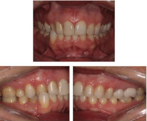
Figure 1: Intra oral photographs, front, right and left.
The facial analysis revealed a harmonic profile and a good proportion of the facial thirds (Figure 2). However, we noticed the increase of the buccal corridor showing transverse maxillary deficiency.
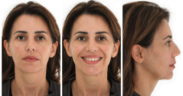
Figure 2: Profile facial analysis, extra oral photographs.
In order to perform the treatment, we chose to make REM (Rapid Maxilla Expansion), with the PC Versatile Mini-implant Figure 3 as an anchorage in the palate, together with the Modified Hyrax apparatus. Choosing the size of the screws by telegraphy and position by the plaster model (Figure 4). In this way, the maxillary bone expansion will occur due to the force exerted directly on the bone, breaking the medial palatine suture, with this technique we avoid orthognathic surgery even in view of the end of growth. For the force is directly bone, not exerting force on the teeth and in the mucosa of the palate, also avoiding the inclination of the molars and necrosis.
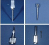
Figure 3: Mini Versatile PC Implant.
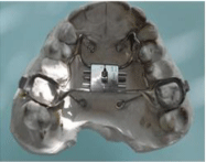
Figure 4: Modified Hyrax apparatus.
After the modified Hyrax was made, the cementation was performed with glass ionomer (Meron) in elements 16 and 26. With Hyrax cementated, the anesthesia is made (Mepivacaine 2%) on the palate in the region where the four self-drilling screws Figure 5 of 7mm. We use the Versatile PC, because it has the reduced thread pitch that allows better bone compression, besides having a single neckless body, avoiding fractures of the Temporary Anchoring Device (Figure 6).

Figure 5: Cementation and Infiltrative Anesthesia in the Palate.

Figure 6: Installation of the 4 mini screw.
The activation of the device is performed for a period of up to 10 days, 2/4 of a day at the end of the activation, we seal the screw with a composite resin Figure 7 and wait 90 days for the healing process. After this period the alignment was aligned with the bonding of the self-ligating In-ovation bracket C (Figure 8).

Figure 7: 2/4 turn activation and screw sealing.
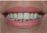
Figure 8: Self Aligned Bracket Alignment.
Finishing the orthodontics with 18 months of treatment we were able to resolve the patient’s main complaint, and improved the smile aesthetics with feldspar porcelain contact lenses, where she chose the natural smile (Figure 9-11).
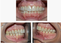
Figure 9: Final treatment result.

Figure 10: Beginning of Treatment.

Figure 11: End of Treatment.
Discussion
The success of maxillary expansion, regardless of the type of breaker, in the treatment of transverse maxillary deficiency is well documented, even in patients with advanced skeletal maturity.
Representing a treatment solution that may prevent surgical intervention. Improving the skeletal effects, aiming to make its proper use the clinical part being operationally feasible.
The mini implants support the force generated by the activation of the expander screw, resulting in the transverse separation of the hemimaxils, which may potentiate the efficiency of orthopedic expansion, as well as facial aesthetics and rehabilitation of the stomatognathic system, with an emphasis on functional stability. orthodontics, seeking an efficient and controllable anchorage to facilitate the movement of natural teeth.
Final Considerations
In the present case, it was possible to verify that after the use of the versatile mini PC implant as an anchorage, there was maxillary expansion without relapses despite the patient’s age chronology. In addition, when compared to conventional, it has the advantage of having a single body, having an intimate bone/screw contact, causing less trauma in the bone tissue minimizing the operative time.
References
- Araújo TM, Nascimento MHA, Bezerra F, Sobral MC. Revista Dental Press de Ortodontia e Ortopedia Facial. Rev. Dental Press Ortodon. Ortop. Facial. Maringá. 2000; 11: 126-156.
- Arismendi JAE. Evaluation of the stability of mini-implants as bone anchoring for upper molar intrusion. Rev Fac Odontol Univ Antioq. 2007; 19: 60-74.
- Bae SM, et al. Clinical application of micro-implant anchorage. J Clin Orthod. 2002; 36: 298-302.
- Bae SM, Kyung HM. Mandibular molar intrusion with miniscrew anchorage. 2006; 40: 107-108.
- Lione R, Franchib L, Cozzac P. Does rapid maxillary expansion induce adverse effects in growing subjects? Angle Orthod. 2013; 83: 172-182.
- Mcnamara JA. Maxillary transverse deficiency. American Journal of Orthodontics and Dentofacial Orthopedics. 2000; 117: 567-570.
- Suzuki H, et al. Miniscrew-assisted rapid palatal expander (MARPE): The quest for pure orthopedic movement. Dental Press J Orthod. 2016; 21: 17-23.