
Special Article - Periodontology
Austin J Dent. 2019; 6(2): 1129.
Management of Bone Defect in Anterior Teeth using Modified Whales Tail Technique - a Case Report
Noble S* and Batra P
¹Department of Periodontics, Vydehi Institute of Dental Sciences and Research Centre, India
*Corresponding author: Sruthy Noble, Department of Periodontics, Vydehi Institute of Dental Sciences and Research Centre, Bangalore, Karnataka, India
Received: March 18, 2019; Accepted: April 10, 2019; Published: April 17, 2019
Abstract
The Modified Whale’s tail technique was performed to obtain maximum interdental papilla fill in the anterior region after placement of Guided tissue regeneration barrier membrane. This study aims to assess the clinical efficacy of this new technique. This case report describes a case with a probing depth of 6-7 mm in the maxillary anterior teeth and their treatment with Modified Whale’s tail technique to obtain regeneration and maximum papilla preservation. The application of the “Whale’s tail” flap lead to significant improvement of hard and soft tissue conditions and improved the clinical attachment level involving the maxillary anterior teeth to recreate a functional and esthetic results.
Keywords: Bone Defect; Periodontics; Whale’s Tail Technique
Introduction
Different surgical procedures have been proposed to preserve the interdental papillary structure during the early and late phases of wound healing to prevent contamination of the regenerating area and subsequent wound failure [1]. These procedures provided greater stability to the blood clot to enhance the regenerative potential [2]. To ensure predictable results in periodontal regeneration, primary closure of the osseous defect is essential to improve clinical attachment gain. To obtain complete coverage of the regenerative material placed in the osseous defect the flap design should be designed in such a way that maximum amount of gingival tissue is preserved [3-5].
To preserve the interdental soft tissue for maximum soft tissue coverage following surgical intervention involving the treatment of proximal osseous defects, Takei et al., proposed a surgical approach called papilla preservation technique [1]. Later Cortellini et al. gave modifications of the flap design - modified papilla preservation flap and simplified papilla preservation flap to be used in combination with regenerative procedures [3].
In 2009, Bianchi and Bassetti described a new surgical technique - the “Whale’s tail” technique, which was designed for the treatment of wide intrabony defects in the esthetic zone. This technique involved the elevation of a large flap from the buccal to the palatal side to facilitate access and visualization of the intrabony defect and was created, especially to perform regeneration while maintaining interdental tissue over grafting material [5]. Authors described a significant improvement on clinical parameters and soft tissue healing with primary closure.
Guided Tissue Regeneration (GTR) comprises procedures attempting to regenerate lost periodontal structures through differential tissue responses.
The aim of this study is to describe a clinical case where the “modified whale’s tail” technique was employed to obtain periodontal regeneration and resulted in gain of clinical attachment level by using barrier membrane in the area of defect.
Case Presentation
A 35 years old woman presenting a diastema between central and lateral right incisors reported to our Department of Periodontics to treat periodontal defects (Figure 1). The patient did not have any systemic problems. There was a clinical and radiographic evidence of an intrabony defect associated with the maxillary right central and lateral incisor with a probing depth of 7 mm and attachment loss of 4mm.
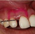
Figure 1:
Before the surgical procedure, Periodontal Probing Depth (PPD), Clinical Attachment Level (CAL) and gingival recessions were recorded. The patient received oral hygiene instructions and full mouth scaling and root planning.
After adequate anesthesia, two semilunar incisions were made from the mucogingival line to the distal margin of the lateral incisor and mesial margin of the central incisor on the buccal surface. In the coronal aspect of the flap, intrasulcular incisions were made at buccal, interproximal and palatal sides. A full-thickness flap was elevated from buccal to palatal side through the diastema (Figure 2,3). The defect was debrided, the root was scaled, planned and conditioned with citric acid for 3 minutes. A bioresorbable bovine collagen membrane was used to cover the defect (Figure 4). The flap was repositioned from the palatal to the buccal side, and its margins were sutured without tension, far away from defect which reduced the chances of “wicking effect” of suture materials (Figure 5,6). Antibiotics were prescribed (Amoxicilin, three times a day) for one week. Healing was uneventful. Recall appointments were performed after 14 days and after a month at the following period (Figure 7). Soft tissue healing depends upon factors such as the surgical technique, flap design, soft tissues management during surgery, root preparation, patient compliance etc.
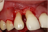
Figure 2:
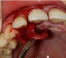
Figure 3:
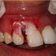
Figure 4:
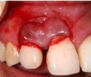
Figure 5:
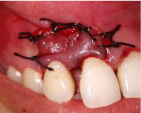
Figure 6:
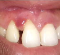
Figure 7:
Discussion
Besides positioning of incisions away from the defect area and placement of sutures distant from the regenerated defects decreased the chances of bacterial colonization of the biomaterials, which is often responsible for regenerative failures.5Bianchi and Bassetti used this technique and showed a mean probing attachment level gain of 4.9 ± 1.8 mm and a probing pocket depth reduction of 5.8 ± 2.5 mm. Our cases showed similar results with a mean probing depth reduction of 3mm and a mean gain in the clinical attachment of 3mm. The main advantages of the technique include good access to the defect area and placement of margins away from the regenerative material, which will prevent the inflammatory response near the regenerative material, thereby increasing the chances of graft uptake. The indications of this technique include therapies aimed at regeneration of periodontal defects such as bone grafts, membrane, or combination of these materials, surgical treatment of anterior tooth region with diastema present. The contraindications include high frenal attachment, recession, and diastema ‹2mm.
Furthermore, the systematic use of incisions distant from the defects and biomaterial margins drastically reduced the percentage of flap dehiscence in this case.
Conclusion
This study suggests that with ideal gingival flap management, enhanced regenerative and esthetic results can be achieved. Application of the “whale’s tail” flap has indeed lead to clinically significant improvement of hard and soft tissue conditions of wide intrabony defects there by creating a functional attachment with esthetic results.
References
- Takei HH, Han TJ, Carranza FA Jr, Kenney EB, Lekovic V. Flap technique for periodontal bone implants. Papilla preservation technique, J. Periodontol. 1985; 56: 204-210.
- Pellegrini G, Pagni G, Rasperini G. Surgical approaches based on biological objectives: GTR versus GBR techniques, Int. J. Dent. 2013; 521-547.
- Cortellini P, Prato GP, Tonetti MS. The modified papilla preservation technique. A new surgical approach for interproximal regenerative procedures. J Periodontol. 1995; 66: 261-266.
- Murphy KG. Interproximal tissue maintenance in GTR procedures: Description of a surgical technique and 1-year reentry results. Int J Periodontics Restorative Dent. 1996; 16: 463-477.
- Bianchi AE, Bassetti A. Flap design for guided tissue regeneration surgery in the esthetic zone: The “Whale’s tail” technique. Int J Periodontics Restorative Dent. 2009; 29: 153-159.