Case Report
Austin J Dermatolog. 2014;1(1): 1003.
An Unusual Manifestation of Cutaneous Sarcoidosis in the Oral Cavity
Firas Al-Niaimi*
Department of Dermatology, Locum positions in UK hospitals, London
*Corresponding author: Firas Al-Niaimi, Locum positions in UK hospitals, Clinic Canary Wharf, 34 N Colonnade, London E14 5HX
Received: March 11, 2014; Accepted: March 26, 2014; Published: March 28, 2014
Introduction
Sarcoidosis is a multi-system, non-caseating granulomatous disease of unknown aetiology [1]. The disease commonly affects the lungs, lymph nodes, skin and eyes. Oral involvement of sarcoidosis is rare [2]. Here we report an unusual oral manifestation in a patient with cutaneous sarcoidosis with complete resolution following methotrexate therapy.
Case Report
34-year old man presented with a 9-month history of painless purple plaques on his face and purple discolouration of his right ear. On further questioning, he also noticed similar looking lesions in his mouth that developed at the same time. He was otherwise systemically well.
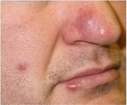
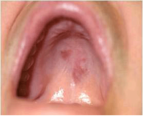
Physical examination revealed a well, afebrile patient with normal vital signs. There was an indurated, purple-red plaque on his nose and right ear, and a purple-red nodule on his right cheek (Figure 1). Oral examination demonstrated a similar nodule on the vermillion border of his lower lip and similar looking purple-red plaque-like lesions on his palate (Figure 2). The lesions were all asymptomatic. An incisional biopsy from a cutaneous lesion showed epithelioid histiocytes forming discrete non-caseating granulomas with minimal lymphocytic infiltrate and no necrobiosis. The features were consistent with sarcoidosis. A biopsy from the palatal lesion showed a similar granulomatous infiltrate consistent with sarcoidosisExamination of other systems was unremarkable. Computed tomography of the chest and abdomen showed no evidence of internal organ involvement or lymphadenopathy. Biochemical investigations showed normal serum calcium and angiotensin-converting enzyme levels. A diagnosis of cutaneous sarcoidosis was made.
Figure 1: Indurated, purple plaque and nodules on the face.
Figure 2: Purple-red plaque-like lesions on the palate.
Initial treatment consisted of potent topical corticosteroids (Clobetasol Propionate 0.05% ointment) which failed to resolve the lesions. Treatment with systemic hydroxychloroquine at a dose of 400 mg/day was similarly ineffective. Subsequently, the patient received oral methotrexate at a dose of 15 mg/week, which resulted in significant improvement in size and discolouration of both the palatal and cutaneous lesions within three months and complete resolution of the palatal lesion in six months. Marked reduction of the palatal lesion was also observed following methotrexate therapy. Computed tomography of the sinuses was unremarkable excluding any granulomas, tumor or sinus pathology that may have extended through the palate.
Discussion
Oral involvement is a rare manifestation of sarcoidosis [2]. The first suspected case of sarcoid granulomas in the oral mucosa was reported in 1942 by Schroff [3]. Oral involvement may be solitary, or part of a generalized cutaneous or systemic sarcoidosis [2,4]. In one third of cases, oral lesions may be the first manifestation of the disease [5].
Clinical presentations of oral mucosal lesions vary and have been reported as painless swellings, nodules, plaque-like lesions and ulcers [2,5]. Various sites of oral sarcoidosis have been reported including the labial, palatal, buccal mucosa, gingival, glossal, floor of mouth sublingual gland, minor salivary glands, submental, and submandibular lymph nodes, maxillary, and mandibular bones [6]. The jaw bones are the most common affected sites of oral involvement in sarcoidosis, whereas the buccal mucosa is the commonest site involved in the soft tissues of the mouth [5].
The diagnosis of oral sarcoidosis is established by clinical features with histologic evidence of non-caseating epithelioid granulomas, and can be supported by biochemical tests and imaging. An important differential diagnosis is orofacial granulomatosis which includes bacterial infections (tuberculosis, syphilis, cat-scatch disease and leprosy), fungal infections (histoplasmosis, coccidiodomycosis), foreign body granulomas and Crohn's disease [2,5].
Management of oral sarcoidosis has been variable, ranging from no treatment to radiation therapy [2]. Surgical excision and curettage was found to be the most common therapy, followed by systemic therapy such as steroids, methotrexate, hydrochloroquine, and minocycline [2,5].
In conclusion, oral involvement in sarcoidosis is rare and may be the solitary or present as an initial manifestation of the disease. Examination of the oral cavity should be performed in every patient with sarcoidosis.
References
- James DG. Descriptive definition and historic aspects of sarcoidosis. Clin Chest Med. 1997; 18: 663-679.
- Suresh L, Radfar L. Oral sarcoidosis: a review of the literature. Oral Dis. 2005; 11: 138-145.
- Schroff J. Sarcoid of the face (Besnier-Boeck-Schumann) disease: report of a case. JADA 1942; 29: 2208-2211.
- Suresh L,Martinez Calixto LE, Aguirre A, Fischman S. Mid-line swelling of the palate. J Laryngol Otol. 2004; 118: 385-387.
- Marie I, Proux A, Levesque H, Bony-Rerolle S, Chenal P. Tongue involvement revealing sarcoidosis. Q J Med 2008; 101: 909-911.

