
Case Report
Austin J Dermatolog. 2014;1(1): 1004.
Dermoscopic Findings in Eruptive Vellus Hair Cysts: A Case Report
Sanami Takada, Yaei Togawa*, Seiichiro Wakabayashi, Naotomo Kambe and Hiroyuki Matsue
Department of Dermatology, Chiba University of Graduate School of Medicine, Japan
*Corresponding author: Yaei Togawa, Department of Dermatology, Chiba University of Graduate School of Medicine, 1-8-1, Inohana, Chuo-ku, Chiba, 260-8670, Japan
Received: March 11, 2014; Accepted: April 07, 2014; Published: April 09, 2014
Abstract
Eruptive vellus hair cysts are a type of cutaneous cyst of skin appendages which is usually seen in children and teenager. We report a case of 7–yearold girl presented with 2 years history of multiple, small, brown or blue–gray papules on her chest. Dermoscopic examination of the papules showed nonfollicular blue homogeneous pigmentation. Histopathologically, the cause of blue homogeneous pigmentation was a cystic structure which was located in the mid–dermis and it had connected to an atrophic hair follicle. We think that non–follicular blue homogeneous pigmentation is one of unique dermoscopic findings to eruptive vellus hair cysts because of its lighter, paler blue color with faded outline.
Keywords: Eruptive vellus hair cysts; Non–follicular blue homogeneous pigmentation; Atrophic hair follicle.
Background
Eruptive vellus hair cysts are a type of cutaneous cyst of skin appendages. They are usually seen in children, adolescents, or young adults and they manifest as smooth papules 1 to 4 mm in diameter, most commonly on the chest. They are benign lesions, and spontaneous resolution occurs in an estimated 25 percent of lesion [1,2], but they may be cosmetically bothersome [3]. One of the dermoscopic findings in eruptive vellus hair cysts located in upper dermis is yellowish white homogenous areas that we have previously reported [4]. We performed dermoscopic examination in a child case of eruptive vellus hair cysts and report another new findings.
Case Presentation
A 7–year–old girl without a past or family history presented with multiple, small, asymptomatic papules localized to her chest. The lesions had been present for approximately 2 years.
Physical examination revealed multiple, 1–2 mm, smooth, brown or blue–gray firm papules on the chest (Figure 1). Dermoscopic examination of the lesions showed non–follicular blue homogeneous pigmentation which had lighter, paler blue color with faded outline (Figure 2).
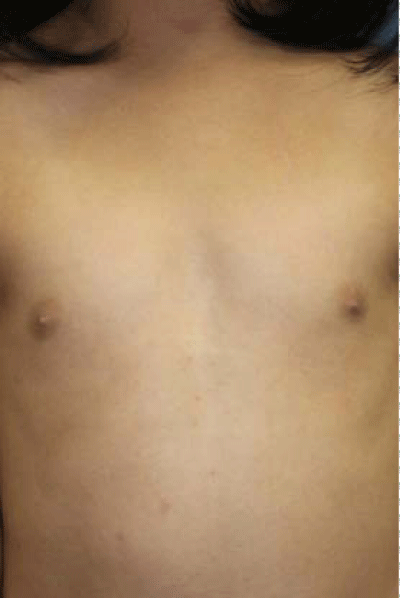
Figure 1: Multiple, 1–2 mm, smooth, brown or blue–gray firm papules on the chest.
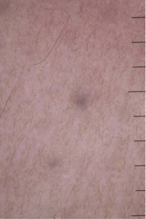
Figure 2: Dermoscopic examination showed non–follicular homogeneous blue pigmentation which had lighter, paler blue color with faded outline. The unit of scale bar is millimeter.
Histopathology revealed that a cystic structure was located in the mid–dermis, and was lined by squamous epithelium. It contained multiple vellus hairs (Figure 3A). In serial section, the cystic structure had connected to an atrophic hair follicle (Figure 3B). Based on these findings, a diagnosis of eruptive vellus hair cysts was established. The patient and her family did not request treatment of the lesions.
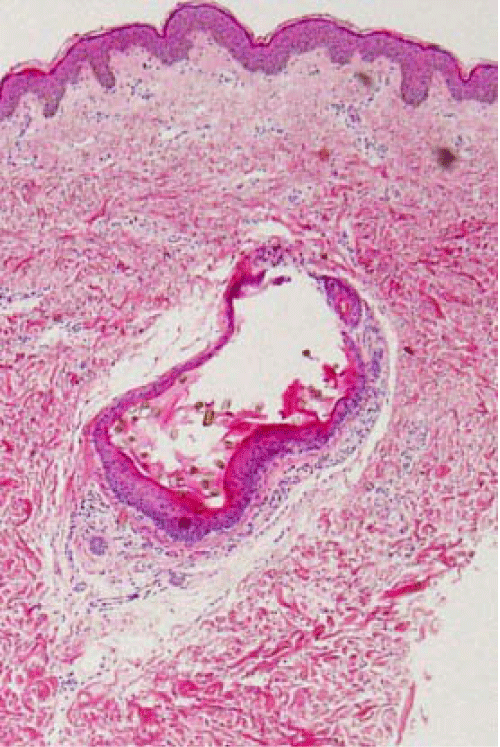
Figure 3A: Histopathological findings. (A) A cystic structure in the middermis was lined by squamous epithelium, and contained multiple vellus hairs (hematoxylin–eosin, original magnification x100).
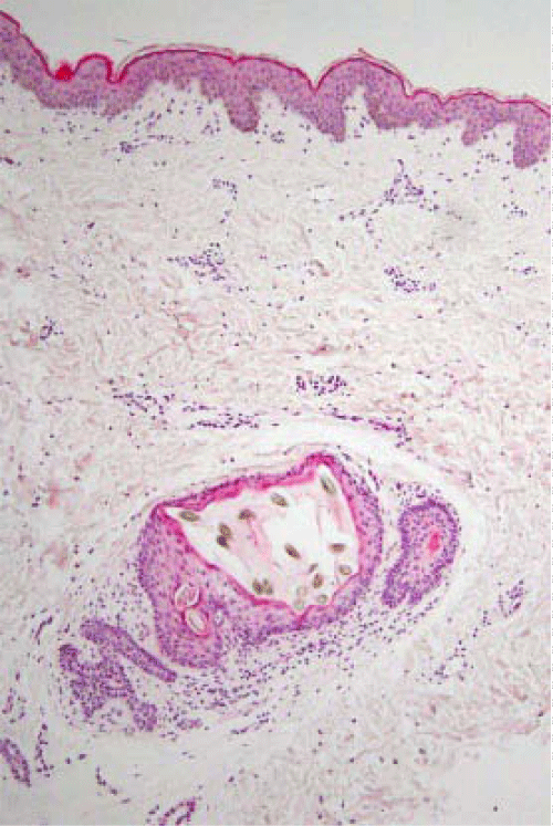
Figure 3B: In serial section, an atrophic follicle had connected to the cyst (hematoxylin–eosin, original magnification x100).
Dermoscopic findings of eruptive vellus hair cysts have not been described without previous our case which was recognized yellowish white homogenous areas [4]. We think yellowish white homogenous areas are common and typical findings of the tumor. However, dermoscopic examination in our case revealed non–follicular homogeneous blue pigmentation unlike the previous case. The cause of this finding was the cysts connected to the atrophic hair follicles in the mid–dermis histologically. In addition, there was no normal hair follicle around the cysts. Many eruptive vellus hair cysts, indeed, have been reported as follicular papules, but some cases have been reported as non–follicular papules like our case clinically [5]. The blue color of our case in dermoscopy showed it located deeper than the superficial “yellowish white” follicular cysts. The finding of non–follicular blue homogeneous pigmentation leaded that the differential diagnosis included blue nevus and hemangioma. However, we thought it was one of unique dermoscopic findings to eruptive vellus hair cysts because of its lighter, paler blue color with faded outline, whereas blue nevi was more darker, grayish blue with sharp demarcation. Moreover, scattered multiple lesions were different from findings of hemangioma such as cavernous hemangioma.
Conclusion
Based on our findings, we conclude that non–follicular blue homogeneous pigmentation is one of characteristic dermoscopic findings of eruptive vellus hair cysts.
Acknowledgement
We are grateful to Prof. Masaru Tanaka (Tokyo Women's Medical University, Medical Center East,Tokyo, Japan) for comments on the dermoscopic findings.
Conflicts of interest
The authors have no conflicts of interest to declare.
References
- Bovenmyer DA. Eruptive vellus hair cysts. Arch Dermatol. 1979; 115: 338-339.
- Sina B, Burnett JW. Eruptive vellus hair cysts. Cutis. 1984; 33: 503-504.
- Lee S, Kim JG. Eruptive vellus hair cyst clinical and histologic findings. Arch Dermatol. 1979; 115: 744-746.
- Togawa Y, Kambe N, Kamada N, Matsue H. Study of 30 cases of nodular lesions presenting with dermoscopy on yellowish. The 111th Annual Meeting of the Japanese Dermatological Association. June 1-3. 2012. Kyoto, Japan.
- Sakamoto F, Shidara A, Satou Y. Eruptive vellus hair cysts. Jpn J Dermatol. 1981; 91: 911-916.