Case Presentation
Austin J Dermatolog. 2014;1(2): 1010.
Psoriasiform Contact Dermatitis Responding to High-Dose Folic Acid
Peter J Aronson* and Jinmeng Zhang
Department of Dermatology, Wayne State University, USA
*Corresponding author: Peter J Aronson, Department of Dermatology, Wayne State University, Dingell Veterans Administration Medical Center, Dermatology 11-M DERM, 4646 John R, Detroit, MI 48201, USA
Received: April 27, 2014; Accepted: June 06, 2014; Published: June 10, 2014
Abstract
We report a case of allergic contact dermatitis in a patch tested area with histopathologic examination showing psoriasiform dermatitis with that and another area with the same histopathology clearing using only daily oral doses of 6 mg folic acid, 100 mg vitamin B6 and 100 mcg vitamin B12. We found a negative correlation coefficient of -0.82 in aggregate for all psoriasiform dermatitis areas proven by biopsy. On the same patient other areas when biopsied showed lichen simplex chronicus. These areas in aggregate showed negative correlation of body surface area involved of only -0.52 despite treatment including not only the administration of these vitamins but also intralesional and topical corticosteroids and other topical immune modulators. High dose folic acid appears to be a potential therapy for patients with allergic contact dermatitis though this may only be appropriate in patients with high BMI and with comorbid signs of psoriasis.
Keywords: Contact dermatitis; Psoriasiform dermatitis; Folic acid; Leptin
Abbreviations
BSA: Body Surface Area; eNOS: Endothelial Nitric Oxide Synthase; LSC: Lichen Simplex Chronicus; iNOS: Inducible Nitric Oxide Synthase
Background
Folic acid is an important vitamin found in many food groups. In high doses it has been demonstrated to have an anti-inflammatory role in psoriasis. Here, we report the first case of psoriasiform contact dermatitis resistant to the standard treatment, yet responding to high-dose folic acid plus vitamins B6 and B12. We propose a possible mechanism based on literature support.
Case Presentation
A 78-year-old man (BMI=33.1) presented with an 8-year history of dyschromic plaques on feet, hands, thighs and hypomelanotic elbow plaques. Previous biopsies in 2005 showed sub acute dermatitis. He also noticed using naftifine gel caused him blisters.
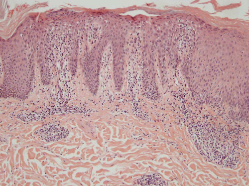
Patch testing performed with the North American Standard 65-allergen series at 72 hours showed reactions to 8 allergens including propylene glycol and formaldehyde. Post testing he showed partial response to allergen avoidance, narrow band-UVB light and intralesional triamcinolone. Using topical clobetasol with propylene glycol and wearing formaldehyde-rich undergarments caused him flares. New scaly lichenified lesions and plaques developed on his legs, hips, buttocks, and at the patch testing site. Five months later, the lesions still remained. Biopsies of the right calf and the patch tested Area of the back demonstrated psoriasiform dermatitis (Figure 1). His vitamin B12 level was normal, and homocysteine level was 13.8umol/L (normal 5-15 μmol/L). He was prescribed daily folic acid 5 mg, vitamin B12 1 mg and B6 100 mg. However, initially the patient did not consistently ingest the full dose of folic acid due to pruritus caused by the vitamins. However after a 4 month-course, his homocysteine decreased to 10.2umol/L. He was also on 2 -10 day courses of prednisone 40 mg daily, which resulted in some plaque flattening. Topical clobetasol 0.05% spray (Galderma®) was prescribed, but after 2 months the rash still covered 2.4% of his Body Surface Area (BSA), with 1.7% on his back. Methotrexate was considered but interferon-γ release assay for tuberculosis (QuantiFERON®-TB Gold Test, Cellestis®) was positive.
Figure 1: Histological specimen of the back lesion: psoriasiform dermatitis (Hematoxylin and Eosin stain 10X).
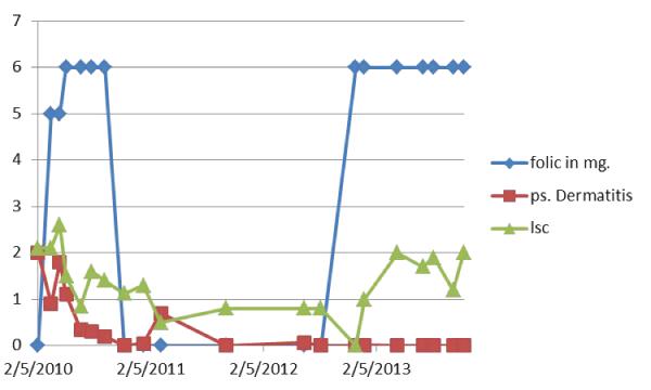
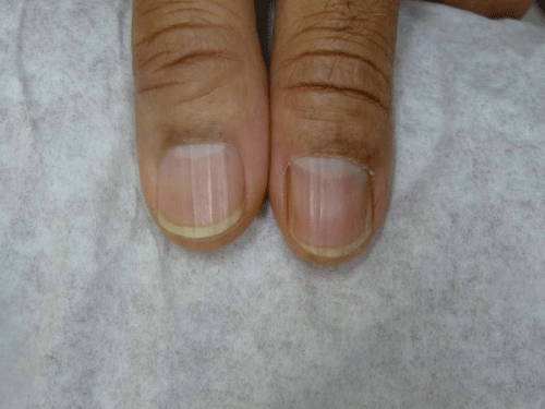
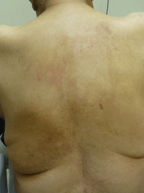
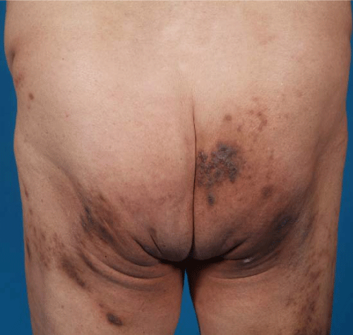
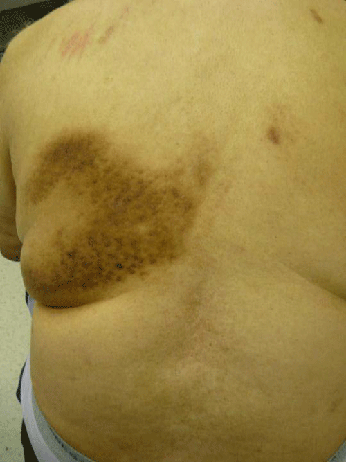
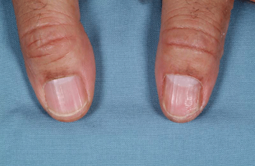
Subsequently (2/05/2010), the involved BSA increased to 4.1% with his back only 0.01% BSA covered but his right calf 2%. His homocysteine was 14.4umol/L., so we restarted the daily regimen of folic acid 5 mg daily, vitamin B6 100mg, and B12 1 mg without any topical treatments. He was noted to have nailed pitting and distal onycholysis in one thumbnail (Figure 2) and minimal pitting on another finger nail. Two months later however the involved BSA had overall increased to 4.4% with the back area newly flaring at 1.8%. His right calf lesion had vanished. His folic acid dose was increased to 6 mg daily. Three weeks later (5/7/2010) the back showed at best some mild flattening (Figure 3), his buttock lesion had not changed (Figure 4) and his calf lesion had reappeared covering 0.35% BSA with total BSA 2.6%. Intralesional triamcinolone and clobetasol spray were then resumed except to the back plaque. After 2 more months of this therapy (6/25/2010) his back was clear and remained clear as of 3-7- 2014 except for some focal erythema in the back location (Figure 5). As of 6/25/2010 his right calf involvement was 0.35% and total BSA 1.2%. By 11/12/2010 his calf cleared only to reappear 1/13/2011. This area was rebiopsied on 4/15/2011 but his time histopathology was not psoriasiform dermatitis but rather Lichen Simplex Chronicus (LSC) as were biopsied lesions on his left calf and buttock. Importantly his nail pitting disappeared with folic acid therapy (Figure 6). Charting comparing folic acid dose with percent BSA covered by biopsy proven psoriasiform dermatitis and LSC showed a possible relationship with folic acid dose and improvement of the former but not the latter (Figure 7). Correlation Coefficient when starting or continuing on 6 mg folic acid for his biopsy proven psoriasiform dermatitis was -0.82 while the correlation coefficient when the patient was on continuing on 6 mg along with topical and intralesional therapies for LSC was less negative at -0.52 . Correlating BSA of just the psoriasiform dermatitis biopsied areas with this patient begun on either 5 or 6 mg. folic acid was not impressive (correlation coefficient was -0.41).
Figure 2: Thumbnails, showing pitting.
Figure 3: Lesion from patch testing site.
Figure 4: Buttock lesion. LSC on biopsy.
Figure 5: Back cleared on vitamins.
Figure 6: Thumbnails showing no pitting.
Figure 7: Percent BSA of areas showing psoriasiform dermatitis and percent BSA of other areas that when biopsied showed LSC compared to daily folic acid dose.
Inducible Isoform of Nitric Oxide Synthase (iNOS) has been implicated in inflammatory skin diseases, specifically contact dermatitis [1]. Folic acid has been found to exert anti-inflammatory effects in vivo and decreases mRNA of iNOS [2]. Dihydrofolate reductase in the folic acid metabolism recycles dihydrobiopterin to tetrahydrobiopterin, which helps stabilize another NOS, dimeric endothelial NOS (dimeric eNOS), reducing oxidative tissue injury. eNOS dimers aid in the catalytic activity of NOS whereas a change from the dimeric form to the inactive monomer of eNOS results in a reduction of net NO synthesis and increases superoxide generation [3]. Therefore, dimeric eNOS is anti-inflammatory, and monomeric is pro-inflammatory [3,4]. Theoretically this suggests that high-dose folic acid taken with vitamins B6 and B12 can have important anti-inflammatory potentials [4,5].
Weight can and may be essential in the modulation of this process. Leptin, a protein released by adipose tissue, has been demonstrated to directly activate NO production in vessels by inducing eNOS phosphorylation [6]. Therefore, with higher leptin in overweight individuals, such as in our patient, more eNOS will be activated. With high-dose folic acid, one theoretically might expect more generation of restored eNOS dimers thus favoring the anti-inflammatory state. Our unusual case suggests that high-dose folic acid can improve contact dermatitis, possibly through its anti-inflammatory role associated with the dimeric nitric oxide synthase and leptin. Importantly lower intake of folic acid might in contrast produce proinflammatory monomeric eNOS and act to flare disease.
Because this man had elbow plaques and one finger had distal onycholysis, nail pitting and other nails had leukonychial spots, the possibility of occult comorbid psoriasis may play a role in this patient's improvement. The lead author has published two case reports of alcoholics with psoriasis responding to high dose folic acid, B6 and B12 [7]. There is sufficient experimental evidence to classify psoriasis and allergic contact dermatitis amongst other inflammatory skin disorders as IL-17 associated diseases [8]. "Inhibition of NOS2 signaling abolishes the de novo induction of Th17 cells and selectively suppresses IL-17 production by established Th17 cells [9]. "In one study of 110 psoriasis patients 47 (42.7%) showed reactions to at least one antigen [10]. However in another publication, an inverse association was found between a psoriasis diagnosis and a positive patch test in two studies [11].
Conclusion
It is premature to generalize this outcome as a potential therapy to all patients with allergic contact dermatitis. We suggest high dose folic acid (6 mg daily), vitamins B6 and B12 as a subject for investigation on obese patients with and without some signs of comorbid psoriasis.
Consent
This case, including proper informed consent form, was reviewed for retrospective case review by the John D. Dingell VA Clinical Investigations Committee and then by the Wayne State University Institutional Review Board. The subject signed the Informed Consent acceptable to the John D. Dingell VA Medical Center, Detroit and to the Wayne State IRB. The authors do not believe the clinical photographs submitted show features that would identify the subject.
References
- Vital AL, Gonçalo M, Cruz MT, Figueiredo A, Duarte CB, Lopes MC. Dexamethasone prevents granulocyte-macrophage colony-stimulating factor-induced nuclear factor-kappa B activation, inducible nitric oxide synthase expression and nitric oxide production in a skin dendritic cell line. Mediators Inflamm. 2003; 12: 71-78.
- Feng D, Zhou Y, Xia M, Ma J. Folic acid inhibits lipopolysaccharide-induced inflammatory response in RAW264.7 macrophages by suppressing MAPKs and NF-κB activation. Inflamm Res. 2011; 60: 817-822.
- Schmidt TS, Alp NJ. Mechanisms for the role of tetrahydrobiopterin in endothelial function and vascular disease. Clin Sci (Lond). 2007; 113: 47-63.
- McCarty MF, Barroso-Aranda J, Contreras F. High-dose folate and dietary purines promote scavenging of peroxynitrite-derived radicals--clinical potential in inflammatory disorders. Med Hypotheses. 2009; 73: 824-834.
- Moat SJ, Madhavan A, Taylor SY, Payne N, Allen RH, Stabler SP, et al. High- but not low-dose folic acid improves endothelial function in coronary artery disease. Eur J Clin Invest. 2006; 36: 850-859.
- Vecchione C, Maffei A, Colella S, Aretini A, Poulet R, Frati G, et al. Leptin effect on endothelial nitric oxide is mediated through Akt-endothelial nitric oxide synthase phosphorylation pathway. Diabetes. 2002; 51: 168-173.
- Aronson PJ, Malick F. Towards rational treatment of severe psoriasis in alcoholics: report of two cases. J Drugs Dermatol. 2010; 9: 405-408.
- Peiser M. Role of Th17 cells in skin inflammation of allergic contact dermatitis. Clin Dev Immunol. 2013; 2013: 261037.
- Obermajer N, Wong JL, Edwards RP, Chen K, Scott M, Khader S, et al. Induction and stability of human Th17 cells require endogenous NOS2 and cGMP-dependent NO signaling. J Exp Med. 2013; 210: 1433-1445.
- Krupashankar DS, Manivasagam SR. Prevalence and relevance of secondary contact sensitizers in subjects with psoriasis. Indian Dermatol Online J. 2012; 3: 177-181.
- Bangsgaard N, Engkilde K, Thyssen JP, Linneberg A, Nielsen NH, Menné T, et al. Inverse relationship between contact allergy and psoriasis: results from a patient- and a population-based study. Br J Dermatol. 2009; 161: 1119-1123.
