
Case Report
Austin J Dermatolog. 2014;1(4): 1017.
Subcutaneous Fat Necrosis of the Newborn: Clinical Manifestations in Four Cases
Tang Jianping* and Shu ye
Department of dermatology, Hunan Children's Hospital, China
*Corresponding author: Tang Jianping, Department of dermatology, Hunan Children's Hospital, 86# Ziyuan Road, Changsha, Hunan, China
Received: May 27, 2014; Accepted: August 18, 2014; Published: August 21, 2014
Abstract
Subcutaneous fat necrosis of the newborn (SFN) is a rare benign neonatal disease of subcutaneous fat tissue, with few cases described in medical literature. The disease affects newborns at term or post-term, with normal general health. We describe four cases, 1 male and 3 female. They all had histories of asphyxia at birth. One of them presented lesions at birth. Three cases appeared skin lesions in 7 to 12 days after birth. The clinical manifestations included multiple indurate, erythematosus plaques and nodules on the back, shoulders, upper arms, buttocks and upper thighs. The lesions were tender with clear edge, smooth surface. Unlike reported in the literature, there were hypocalcaemia in 3 patients.
Keywords: Subcutaneous fat necrosis; Newborn
Introduction
Subcutaneous fat necrosis of the newborn (SFN) is an uncommon disorder characterized by firm, erythematosus nodules and plaques over the trunk, arms, buttocks, thighs, and cheeks of newborns. The nodules and plaques appear in the first several weeks of life. SFN usually runs a self-limited course, but it may be complicated by hypocalcaemia and other metabolic abnormalities. The best recognized risk factors for the development of SFN are asphyxia and hypothermia. We discuss four cases of SFN at term or post-term that developed painful subcutaneous nodules.
Case Presentation
Case 1
A 39-day-old female patient, born at post-term by C section, with severe asphyxia, hypoxia-ischemic encephalopathy and aspiration pneumonia. Twelve days after birth, the patient presented tender nodules on the back and gradually emerged to upper arms, thighs and posterior of neck. Physical examination revealed multiple subcutaneous nodules on the posterior of neck, back, upper arms, buttocks and upper thighs, symmetrically. The margins were well defined, the texture was hard, the surface was smooth, and nodules were tender and movable (Figure 1). Biopsy of the skin lesion was characterized by focal subcutaneous fat tissues necrosis, needle-like cracks in a radial arrangement and a few lymphocytes and histiocytes infiltration (Figure 2-4). Because patient had hypoxia-ischemia encephalopathy, she was given hyperbaric oxygen treatment. We observed that the nodule significantly shrank after hyperbaric oxygen treatment.
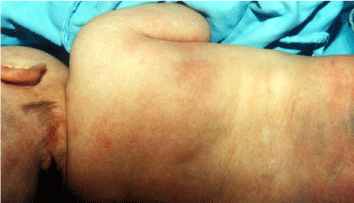
Figure 1: A 39-day-old female patient multiple indurate plaques and nodules on the posterior of neck, back, upper arms, buttocks and upper thighs.
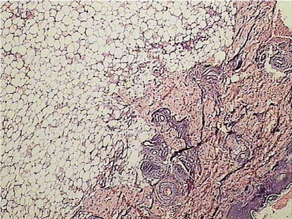
Figure 2: Focal subcutaneous fat tissues necrosis with a few lymphocytes and histocytes in filtration, HE10x4.
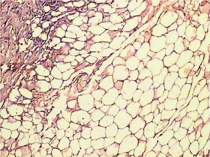
Figure 3: Focal subcutaneous fat tissues necrosis with a few lymphocytes and histocytes infiltration, HE10x10.
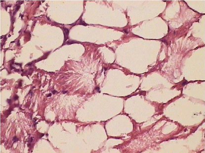
Figure 4: Focal subcutaneous fat tissues necrosis, needle-like cracks in a radiant arrangement, HE10x40.
Case 2
A 41-day-old female patient, born at post-term, with asphyxia. One week after birth, the patient presented red nodules on the bilateral buttocks. New nodules gradually appeared on the back and upper arms. Physical examination revealed multiple subcutaneous indurate nodules of uneven size on upper back, upper arms and buttocks (Figure 5). The lesions were tender with well-defined margins, and smooth surface. Liver was palpated 4cm under ribs, while spleen was palpated 1cm under ribs, and their texture were medium without pain. Blood triglyceride was 2.36mol/L (normal value 0.56-1.7 mmol/L). Blood cholesterol was normal. Blood calcium was 1.92mmol/L (normal value 2.25-2.75 mmol/L). A biopsy of the lesion on back was carried out, showing focal subcutaneous fat tissues necrosis, needle-like cracks in a radial arrangement and infiltration of a few lymphocytes, histiocytes and fibroblasts (Figure 6-8). After admission, the patient was given salvia miltiorrhiza, low molecule dextrin, Vit E, and was kept warm and also received ultra red rays. Subcutaneous nodules shrank significantly, liver enlargement was alleviated and spleen recovered. The subcutaneous nodules disappeared after 3 months.
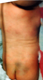
Figure 5: A 41-day-old female patient multiple subcutaneous indurate nodules of uneven size on back, upper arms and buttocks.
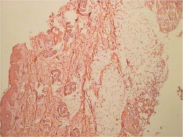
Figure 6: Focal subcutaneous fat tissues necrosis with a few lymphocytes, histiocytes and fibroblasts infiltration, HE10x4.
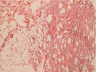
Figure 7: Focal subcutaneous fat tissues necrosis with a few lymphocytes, histiocytes and fibroblasts infiltration, HE10x10.
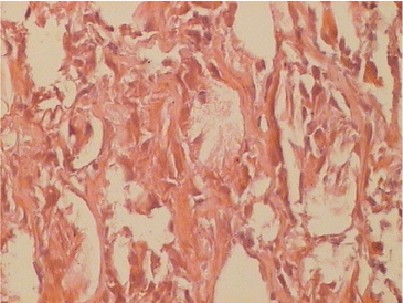
Figure 8: Focal subcutaneous fat tissues necrosis, needle-like cracks in a radiant.
Case 3
A 18-day-old female patient, born at term by C section, with asphyxia. At birth, he presented hardened, erythematosus, edematous lesions on the bilateral buttocks, and gradually enlarged and extended downward without fever. Physical examination revealed firm, erythematosus nodules and plaques from bilateral buttocks to posterior upper thigh. Skin biopsy demonstrated subcutaneous fat tissues necrosis. Blood calcium was 1.14mmol/L. Cranial B-type ultrasound showed choroid plexus hemorrhage (absorption period). Follow-up study found skin nodules disappeared after 2 months.
Case 4
A 2-day-old male patient, born at term by C section, with asphyxia, who admitted to hospital because of tachypnea for 52 hours, his diagnosis was aspiration syndrome with hypoxic ischemic encephalopathy. After admission, patient was given reducing the intracranial pressure, anti-infection and protecting brain treatment and his condition improved markedly. 10 days after admission, the patient presented hardened nodules on the back and gradually emerged to shoulders, buttocks and thighs. Blood calcium was 0.62 mmol/L. Blood lipid level was normal. Cardiac color ultra sonography demonstrated right atrial and ventricular enlargement, acleistocardia and normal heart function. Cranial B-type ultrasound suggested hypoxic ischemic encephalopathy. Skin biopsy of the lesion demonstrated subcutaneous fat tissues necrosis. The patient was given salvia miltiorrhiza, low molecule dextrin, Vit E, and was kept warm and received ultra red rays. After treatment, nodule shrank and softens. He recovered completely from the lesions in the fourth month.
Discussion
In 1933, Cruse firstly reported a case diagnosed as neonatal subcutaneous fat necrosis. It is a rare benign neonatal disease of subcutaneous fat tissue. The typical skin lesions are red or mauve nodules and plaques. The borders are well demarcated, and the texture is rubber or hard. The lesions are asymptomatic and movable, tender in acute stage, and appear on the buttocks, back, limbs or cheeks. Skin lesion biopsy feature are characterized by varying degrees of subcutaneous fat cells degeneration and necrosis with lymphocytes, histiocytes and giant cells infiltration, typical cells having needle-shaped clefts in a radial arrangement. The prognosis is good. Nodules soften and are absorbed without any mark after several months [1]. The complication is hypocalcaemia; SFN may induce severe hypocalcaemia without any visible cutaneous lesion [2]. The cause of SFN is not known. Maybe involved with birth trauma, suffocation and mothers diabetes. Recently subcutaneous fat necrosis has been reported as an unexpected outcome of therapeutic cooling for hypoxic-ischemic encephalopathy [3].
We reported four patients who had suffocation at birth. The onset of 3 cases was within 2 weeks after birth, one of them presented lesions at birth. The general situations were good. Three out of 4 patients were born by C section, 1 patient had intracranial hemorrhage, and 2 patients had hypoxic ischemic encephalopathy. These illustrate that SFN associates with abnormal history of birth and suffocation. It is stated that the complication of SFN is hypocalcaemia, but 3 in 4 cases had hypocalcaemia. The cause is not known and may be related to eating habits difference or calcium deposition in organs and tissues. Four patients were given salvia miltiorrhiza for improving blood circulation. Salvia miltiorrhiza is a traditional Chinese medicine used for circulatory and heart health. It has shown efficacy in treating SFN. Hyperbaric oxygen therapy succeeded for the therapy of 1 case of neonatal subcutaneous fat necrosis, and presents as an alternative to treat SFN patients with hypoxia-ischemia encephalopathy.
References
- Mahé E, Girszyn N, Hadj-Rabia S, Bodemer C, Hamel-Teillac D, De Prost Y. Subcutaneous fat necrosis of the newborn: a systematic evaluation of risk factors, clinical manifestations, complications and outcome of 16 children. Br J Dermatol. 2007; 156: 709-715.
- Bonnemains L, Rouleau S, Sing G, Bouderlique C, Coutant R. Severe neonatal hypercalcemia caused by subcutaneous fat necrosis without any apparent cutaneous lesion. Eur J Pediatr. 2008; 167: 1459-1461.
- Scheans P. Subcutaneous fat necrosis: a complication of neuroprotective cooling. Neonatal Netw. 2012; 31: 409-412.