Case Report
Austin J Dermatolog. 2014;1(5): 1023.
Persistant Urticarial Plaques of Lupus Erythematosus Tumidus: A Case Report
Uzuncakmak TK1*, Karadag AS1, Akdeniz N1, Zindanci I1, Zemheri E2 and Ozkanli S2
1Department of Dermatology, Istanbul Medeniyet University, Turkey
2Department of Pathology, Istanbul Medeniyet University, Turkey
*Corresponding author: Tugba Kevser Uzuncakmak, Department of Dermatology, Istanbul Medeniyet University, Goztepe Research and Training Hospital, Istanbul, Turkey
Received: August 21, 2014; Accepted: September 30, 2014; Published: October 02, 2014
Abstract
Lupus erythematosus tumidus is an uncommon variant of chronic cutaneous lupus erythematosus and characterized by erythematous, pruritic urticarial papules and plaques without any epidermal changes. Lesions are usually localized on sun-exposed areas like face or trunk and commonly resolve without scarring.
We want to present a 43 year-old female patient with pruritic, erythematous and edematous papules and plaques on her back for 8 years. Her lesions first appeared in spring and summer time in first 3 years then remained stable for all seasons. Histopathological examination of these lesions were consistent with lupus erythematosus tumidus and responsed very-well to systemic hydroxychloroquine therapy in one month. We want to remind lupus erythematosus tumidus in differential diagnosis of pruritic, urticarial papules and plaques which are localized on places that do not expose to sun.
Keywords: Cutaneous lupus erythematosus; Lupus erythematosus tumidus; Pruritus
Introduction
Lupus Erythematosus Tumidus (LET) is the most photosensitive variant of cutaneous Lupus Erythematosus (LE) and in clinical practice it is one of the rare variants of LE. Clinical manifestation of LET is characterized by smooth, erythematous urticarial plaques on sun-exposed areas without any epidermal changes, such as erosion, follicular plugs, atrophy or scale against to other forms of lupus erythematosus. Central healing is another feature of LET. Jessner’s lymphocytic infiltration, polymorphous light eruption, Reticular Erythematous Mucinosis (REM), urticarial vasculitis and pseudolymphoma are most important diseases in differential diagnosis [1-3]. Recently LET is offered to be classified as a separate entity more than a subytpe of chronic cutaneous lupus erythematosus as well the presence of lupus tumidus lesions in patients with other types of LE leads to classification as a subtype of LE [4].
Herein we present a female patient with urticarial papules and plaques on her back which was consistent with lupus erythematosus tumidus clinically and histopathologically.
Case Report
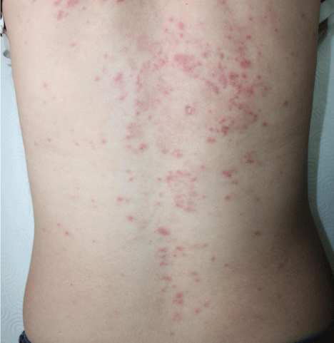
A 48-year-old female patient presented to our outpatient clinic with persistant pruritic erythematous papules and plaques on her back for 8 years (Figure 1). Her lesions appeared in spring and summer time in first three years but then these lesions have persisted for all seasons. She does not have any systemic symptom or a drug history. A punch biopsy and a direct immunfloresence specimen were taken from her back with initial diagnoses of subacute cutaneous lupus erythematosus, sarcoidosis, Grover’s disease, erythema annulare centrifigum, granuloma anulare and polymorphous light eruption from her back.
Figure 1: Erythematous urticarial papules and plaques on back.
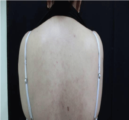
Histopathologic examination revealed basketwave keratosis, mild achantosis in epidermis, lymphocytes, plasma cells and histiocytes infiltration in perivascular and periadnexial areas, and extra-cellular mucin deposits in dermis. (Figure 2). Her direct immunfloresance was negative. In laboratory examination complete blood count, biochemistry, erytrocyte sedimentation rate, anti nuclear antibody, anti ds-DNA, C3, C4 levels, VDRL and TPHA serology were totally normal. She was accepted as lupus erythematosus tumidus with her clinical presentation and laboratory findings. Topical tacrolimus % 0.1 ointment, sunscreens and systemic hydroxychloroquine 400 mg/d and systemic antihistaminic were initiated. Her lesions were totally regressed in one month (Figure 3).
Figure 2: Basketwave keratosis, mild achantosis in epidermis, mononuclear cell infiltration in perivascular and periadnexial areas, and extra-cellular mucin deposits (arrow) in dermis.
Figure 3: Almost totally remission is seen on back.
Discussion
LET was first described by German dermatologist E. Hoffmann in 1909 and reported as a case report by Gougerot and Burnier in 1930 [1]. Prevelance or incidence of this disease is unknown [1]. Age distribution is predicted to be same with CLE and both gender are equally affected [1].This form of LE is recently considered to be a photodermatosis, not a subtype of CLE because of the absence of antibodies, systemic criteria of lupus erythematosus and interface dermatitis in histopathology[1,4]. Our case was supporting this idea with autoantibody negativity, histopathological findings and absence of systemic features of systemic LE.
LET presents with erythematous, urticarial plaques without a surface change as follicular plugging, squam, atrophy, scatris or pigmentation clinically as our patient. Characteristic lesions are usually localized on sun exposed areas as face, shoulders and arms but appearance on non sun exposed areas like buttocks have been reported in the literature [5]. These lesions may appear in 24 hours or several weeks after sun exposure and can persist for weeks or months [3]. In our patient urticarial plaques started with sun exposure at the begining but then remained stable on lomber area which does not expose to sun and persist for years on same localization. LET can be triggered by medications such as biologic agents [6-8]. This reaction may lead misdiagnosis of lupus tumidus with drug-induced LE. Patients with LET are typically ANA negative and rarely display other clinical features of LE as our patient [3,9].
Mucin deposition is a strong marker of this entity especially in differential diagnosis of LET from Jessner’s lymphocytic infiltration. Both perivascular lymphocyte infiltration and mucin deposition are seen in REM and LET but superficial lymphocytes, superficial mucin deposition and less complement and immunglobulin deposition along the dermoepidermal junction are important findings in differential diagnosis of REM from LET histologically [10]. More dense lymphocytic infiltration that can mimic T or B cell lymphoma is expected in pseudolymphoma.
Significant papillary dermal edema is an important finding of polymorphous light eruption, also epidermal spongiosis, eosinophils and neutrophils are other histological findings of polymorhous light eruption [11]. In our patient we did not see any epidermal changes or a dermal dense inflammatory infiltration histopathologically.
Sytemic corticosteroids, antimalarials, topical steroids and tacrolimus are treatment options in LET [2]. Photodynamic therapy and pulse dye laser are also reported to be effective in LET treatment [2,12,13]. Our patient responsed very well to systemic hydroxycloroquine and topical tacrolimus in one month duration.
We want to remind this uncommon variant of LE in differential diagnosis of persistant pruritic urticarial papules and plaques which could be seen on places which do not expose to sun.
References
- Rodríguez-Caruncho C, Bielsa I. [Lupus erythematosus tumidus: a clinical entity still being defined]. Actas Dermosifiliogr. 2011; 102: 668-674.
- Verma P, Sharma S, Yadav P, Namdeo C, Mahajan G. Tumid lupus erythematosus: an intriguing dermatopathological connotation treated successfully with topical tacrolimus and hydroxyxhloroquine combination. Indian J Dermatol. 2014; 59: 210.
- Kuhn A, Richter-Hintz D, Oslislo C, Ruzicka T, Megahed M, Lehmann P. Lupus erythematosus tumidus--a neglected subset of cutaneous Lupus erythematosus: report of 40 cases. Arch Dermatol. 2000; 136: 1033-1041.
- Mutasim DF. Lupus erythematosus tumidus as a separate subtype of cutaneous lupus erythematosus. Br J Dermatol. 2010; 163: 230.
- Nishiyama M, Kanazawa N, Hiroi A, Furukawa F. Lupus erythematosus tumidus in Japan: a case report and a review of the literature. Mod Rheumatol. 2009; 19: 567-572.
- Sohl S, Renner R, Winter U, Bodendorf M, Paasch U, Simon JC, et al. [Drug-induced lupus erythematosus tumidus during treatment with adalimumab]. Hautarzt. 2009; 60: 826-829.
- Schneider SW, Staender S, Schlüter B, Luger TA, Bonsmann G. Infliximab-induced lupus erythematosus tumidus in a patient with rheumatoid arthritis. Arch Dermatol. 2006; 142: 115-116.
- Böckle BC, Baltaci M, Weyrer W, Sepp NT. Bortezomib-induced lupus erythematosus tumidus. Oncologist. 2009; 14: 637-639.
- Walling HW, Sontheimer RD. Cutaneous lupus erythematosus: issues in diagnosis and treatment. Am J Clin Dermatol. 2009; 10: 365-381.
- Cinotti E, Merlo V, Kempf W, Carli C, Kanitakis J, Parodi A, et al. Reticular erythematous mucinosis: histopathological and immunohistochemical features of 25 patients compared with 25 cases of lupus erythematosus tumidus. J Eur Acad Dermatol Venereol. 2014.
- Lim HW, Hawk LM. Photodermatoses. In Dermatology. 2nd edn. Bolognia JL, Jorizzo JL, Rapini RP, editors Spain: Mosby. 2008: 86; 1333-1336.
- Truchuelo MT, Boixeda P, Alcántara J, Moreno C, de las Heras E, Olasolo PJ. Pulsed dye laser as an excellent choice of treatment for lupus tumidus: a prospective study. J Eur Acad Dermatol Venereol. 2012; 26: 1272-1279.
- Erceg A, de Jong EM, van de Kerkhof PC, Seyger MM. The efficacy of pulsed dye laser treatment for inflammatory skin diseases: a systematic review. J Am Acad Dermatol. 2013; 69: 609-615.


