Case Report
Austin J Dermatolog. 2014;1(5): 1025
A Case of Basal Cell Carcinoma on Cavum Conchae Treated by Carbon Dioxide Laser
Kang Daoxian, Zhou Kaihua*, Zou Qing and Wang Lu
Department of Dermatoverenology, China Aviation Industry 363 hospital, China
*Corresponding author: Zhou Kaihua, Department of Dermatoverenology, China Aviation Industry 363 hospital, Chengdu, China
Received: May 12, 2014; Accepted: October 08, 2014; Published: October 10, 2014
Case Report
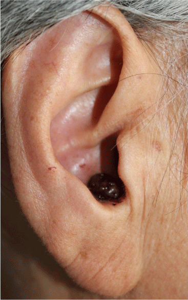
A 67-year old female presented to our clinic with the history of nodule in the right ear for more than a year. A year ago, a dark brown colored millet grain sized nodule appeared on her left ear without any discomfort. She ignored the nodule but it became bigger gradually without any other symptoms. Physical examination revealed a dark brown colored broad bean sized nodule on the right cavum conchae. The surface was relatively smooth and the boundary was clear (Figure 1).
Figure 1: The dark brown colored broad bean sized nodule on the right cavum conchae.
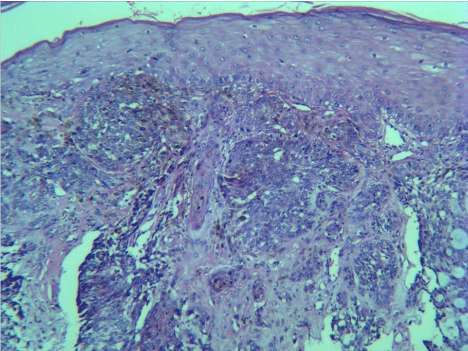
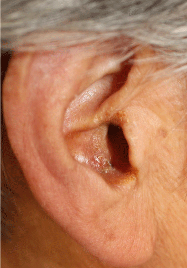
Considering the special location of the lesion and chances of the cartilage atrophy, carbon dioxide laser extraction was chosen rather than surgical removal. After routine disinfection, 2% lidocaine was injected around the nodule for local anesthesia. The power was adjusted to level 3 and the nodule was excised on the base by carbon dioxide laser. Then the base and boundary were burned with additional energy. The removed tissue was prepared for pathological examination. During histopathology examination, it showed that multiple islands of tumor were connected to epidermis made up of basaloid cells with palisading at periphery (Figure 2), which accords with the diagnosis of basal cell carcinoma. Combining with the clinic manifestation, she was diagnosed as pigmented BCC. Twelve days after carbon dioxide laser treatment, the wound healed (Figure 3). There was no suspicious lesion left over. And 5% imiquimod cream was topical used for 2 months. There was no recurrence was observed for 10 months follow-up.
Figure 2: HPE showed multiple islands of tumor were connected to epidermis made up of basaloid cells with palisading at periphery (x100).
Figure 3: The nodule disappeared after treatment for 3 months.
Discussion
Basal Cell Carcinoma (BCC) is almost exclusively seen in head-neck region with rare involvement of trunk and extremities. The tumor is commonly seen on nose, eyelids, at the inner can thus of eyes and behind the ears [1]. In this case, the tumor was present on cavum conchae of the right ear. The special location often leads to misdiagnosis. Besides, it is difficult to perform surgery and apply suture on that anatomical area.
CO2 laser was used to remove the tumor because of its good cutting performance. Moreover, the laser rarely causes bleeding and oozing with their coagulation characteristic [2]. Therefore this procedure doesn't require suturing unlike surgery. Imiquimod is an immune response modifier. There are a lot of data which suggests that imiquimod may be used in the treatment of BCCs [3]. Thus, the 5% imiquimod cream was used as an additional treatment on the base.
In this case, we report a kind of common tumor in an uncommon location, where it's difficult to have an operation. CO2 laser combined with 5% imiquimod cream gives this patient a good resolve.
References
- Tambe SA, Ghate SS, Jerajani HR. Adenoid type of Basal cell carcinoma: rare histopathological variant at an unusual location. Indian J Dermatol. 2013; 58: 159.
- Kim SK, Park JY, Song HS, Kim YS, Kim YC. Photodynamic therapy with ablative carbon dioxide fractional laser for treating Bowen disease. Ann Dermatol. 2013; 25: 335-339.
- Smith V, Walton S. Treatment of facial Basal cell carcinoma: a review. J Skin Cancer. 2011; 380371.


