1Department of Dermatology, Chi Mei Medical Center, Taiwan
2Center for General Education, Southern Taiwan University of Science and Technology, Taiwan
*Corresponding author: Feng-Jie Lai, Department of Dermatology, Chi Mei Medical Center, No.901, Zhonghua Rd, Yongkang Dist, Tainan City 710, Taiwan,
Received: October 15, 2014; Accepted: October 20, 2014; Published:October 22, 2014
Citation: Pai-Shan Cheng and Feng-Jie Lai. Disseminated Superficial Porokeratosis. Austin J Dermatolog. 2014;1(6): 1027. ISSN:2381-9189
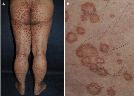
A 48-year-old man presented with a 10-year history of extensive slowly progressing lesions on his trunk, extremities, perineum and face. Physical examination revealed multiple brownish slightly atrophic patches surrounded by elevated, hyperkeratotic borders (Panel A). The lesions all began as a small keratotic papule which spread centrifugally and then atrophied in the center. Therefore, a circinate well-defined plaque which was surrounded by a keratotic wall formed, and gave rise to a festooned appearance (Panel B). On the basis of this characteristic presentation, a diagnosis of disseminated superficial porokeratosis was made. Porokeratosis is comprised of a heterogeneous group of disorders that are inherited in an autosomal dominant fashion. Immunosuppression, radiation therapy and ultraviolet exposure may exacerbate porokeratosis and promote the development of skin cancers. Malignant transformation happens in about 7 to 11 percent of patients, and squamous cell carcinoma is the most common associated malignancy.
Sample Text.
