
Department of Dermatology and Pathology, Istanbul Medeniyet University, Goztepe Research and Training Hospital, Turkey
*Corresponding author: Ayse Serap Karadag, Department of Dermatology, Istanbul Medeniyet University, School of Medicine, Turkey
Received: October 28, 2014; Accepted: October 31, 2014; Published: November 03, 2014
Citation: Karadag AS, Ozlu E and Ozkanli S. Brown-Black, Papular, Verrucous Lesion on the Face: What is Your Diagnosis?. Austin J Dermatolog. 2014;1(6): 1029. ISSN:2381-9189
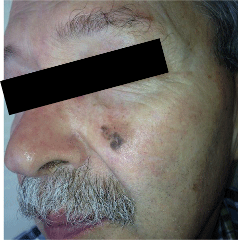
A 70 years old male patient admitted to our clinic with a 10 years history brown, macular lesion that is 1×1,5 cm in size and up on that lesion, a 1 year history, sharp edged, brown- black, verrucose, popular lesion that is protruded from the skin and is 0,5×0,5 cm in size (Figure 1a).
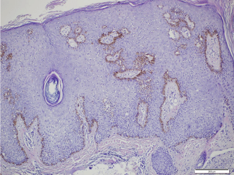
Skin biopsy taken from the lesion revealed hyperkeratosis, acanthosis, scattered melanocytes in the epidermis and lymphocytes and macrophages in the superficial dermis, but didn’t reveal any atypiaormitosis (Figure 1b).
What is your diagnosis?
What is your treatment approach?
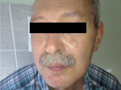
We applied cryotherapy two times, and then the entire lesion cured (Figure 1c).
Melanoacanthoma is a solitary benign skin tumor that is common with middle- old aged people and is usually placed on the head, trunk, lips or eyelids. Melanoacanthoma can imitate the clinical presentations of seborrheic keratosis and malign melanoma. But characteristic histopathological attributes allow differentiation of these 3 diseases. Recently, melanoacanthoma is thought to a rare variant of seborrheic keratosis rather than a distinct entity. Melanoacanthoma is a benign skin tumor, so it can be treated with curettage, cryotherapy or simple excision.
Brown-black, maculopapular lesion that is 1×1,5 cm in size.
Hyperkeratosis, acanthosis, scattered melanocytes in theepidermis and lymphocytes and macrophages in the superficial dermis but didn’t revealanyatypiaormitosis.
Lesion regressed after cryotherapy treatment.
Austin Publishing Group is an emerging open access publisher specialising in Science, Technology and Medicine is dedicated to serve the biomedical community through its initiatives. Austin Publishing Group is an academic publisher with 100+ peer reviewed open access journals in various subjects such as biomedical, Pharma, Life Sciences, Environmental, Engineering and Management. Austin Publishing Group publishes Open Access eBooks providing free access to vast scientific literature.