Case Report
Austin J Dermatolog. 2015;2(1): 1034.
Childhood Granulomatous Perioral Dermatitis with Good Responses to Minocycline and Topical Tacrolimus, Extraordinary Significance
Sami Loai and Changzheng Huang*
Department of Dermatology, Huazhong University of Science and Technology, China
*Corresponding author: Changzheng Huang, Department of Dermatology, Union Hospital, Tongji Medical College, Huazhong University of Science and Technology, Wuhan 430022, China
Received: February 04, 2015; Accepted: February 25, 2015; Published: February 27, 2015
Abstract
Childhood Granulomatous Perioral Dermatitis (CGPD) is a rare disease characterized as an eruption, which is seen predominantly in children and represents a granulomatous form of perioral dermatitis or of acne agminata. It is a condition of unknown etiology affects prepubescent children of both sexes and typically persists in several months, characterized by asymptomatic papular eruptions generally confined to the central area of the face with clustering around the mouth, eyes and ears. The histopathology shows a granulomatous pattern. We report a 24-year-old Chinese girl with multiple, discrete, red to brown papules on erythematous base of 2-months duration on the perioral and periocular areas. Histopathological examination demonstrated dermal granulomatous infiltrates. The patient show good response to the combination treatment of oral Minocycline and topical Tacrolimus. CGPD is the disease of children, but in this rare case the disease occur in adult, with good response to oral Minocycline and topical Tacrolimus.
Keywords: Granuloma; Perioral Dermatitis; Childhood
Introduction
Childhood Granulomatous Perioral Dermatitis (CGPD), also known as Facial Afro-Caribbean Childhood Eruption (FACE), is a distinctive granulomatous form of perioral dermatitis [1]. The histology has been variously described as showing non-specific inflammation with hyperkeratosis or, as granulomatous with the inflammatory changes often, but not invariably, being perifollicular. Complete resolution few months after the appearance of skin lesions, either spontaneously or in response to treatment. Some time with small pitted scars. Systemic treatment has been achieved with erythromycin or metronidazole [2,3].
Case Report
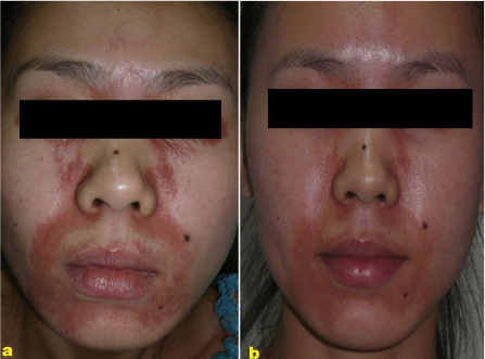
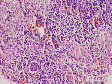
A 24-year-old Chinese girl presented with a Two-month history of pruritic papules on the face. The eruption started on the perioral area with red papules on the erythematous background. After twenty days, the eruption spread upward to involve the side of the nose and periorbital areas include upper and lower eyelids. She had neither past history of skin disorders, nor drug history whether topical or systemic. She was otherwise healthy. No remarkable family history could be found. Laboratory evaluation including routine blood, urinary, liver and kidney function analysis, Purified Protein derivative test, chest X-ray show no significant foundlings. Clinical examination of the patient show multiple small red to brown colored papules on the base of erythematous plaque located on central areas of the face including the perioral area , around the nose and periorbital area with upper and lower eyelids involved (Figure 1a). However, no pustules or papulovesicles were found. A biopsy of facial papules had been done and shows a granulomatous infiltration composed of lymphocytes, histiocytes, epitheliod cells and multinucleated giant cells, but without caseation necrosis in dermis (Figure 2) PA and acid-fast stain was negative. Cultures for fungus were also negative. A final diagnosis of childhood granulomatous perioral dermatitis in adult patients was established based on the clinical manifestation and histopathology. The patient received oral Minocycline 50mg bid and topical Tacrolimus ointment 0.03% twice daily with good responses and resolving of the majority of the skin lesions after 21 days and more improvement after 50 days . Follow up to the patient after 10 weeks shows better improvement include to decrease from the number of the papules and diminish the color of the eruption without observed scars (Figure 1b).

    Figure 1:  CGPD in adult before ‘(a)’ and after ‘(b)’ treatment: ‘(a)’A close up
view shows papules on the red base around the mouth, the side of the nose
and around the eyes. View ‘(b)’ shows dramatic improvement following ten
weeks treatment.

    Figure 2:  Schematic diagram of histopathology on high power, there
were granulomatous infiltration in the dermis composed of lymphocytes,
histiocytes, epitheliod cells and multinucleated giant cells with no caseation
necrosis (HE×400).
Discussion
Perioral dermatitis is a well-recognized Papulopustular facial dermatitis affecting children ranged from 7 months to 13 years and young people especially young women with an average age of onset between 25 and 35 years, the etiology is still unclear. However, uncritical use of topical corticosteroids especially topical fluorinated corticosteroid often precedes skin lesions [4]. Childhood Granulomatous Perioral Dermatitis (CGPD) was first reported on Gianotti F in 1970s [1] described five Italian children aged 2 to 7 years with papules, pustules and diffuse erythema concentrated around the mouth, eyes and nose clinically and granulomatous infiltrate concentrated around the upper dermis and perifollicles histopathologically. Later on , it has been termed as granulomatous perioral dermatitis in children by Frieden et al., FACE by Williams et al. [5,6]. CGPD has been considered as a distinctive granulomatous form of perioral dermatitis or a condition included in the spectrum of rosacea and perioral dermatitis. It usually affects prepubescent children and is common in Dark-skin patients, even though a few cases of Fair-skin children have been reported 4. The skin lesions are usually 1-3 mm, monomorphic, erythematous or hypopigmented, or Yellow-brown or flesh-colored asymptomatic papules and typical characteristic by absence of pustules. The disorder is usually considered as a self-limited disease and heals spontaneously with no scarring formation [5]. Our case is a 24-year-old female. The skin lesions and their distribution and histopathological changes are typical as seen in CGPD.
It has been reported long term topical use of potent steroids may induce or exacerbate CGPD [5]. Other possible factors related to this condition include allergens, irritants, or reactions to bubble gum, formaldehyde, cosmetic preparations and antiseptic solutions or even to varicella vaccination [6]. The histopathological changes include mild Spongiosis and elongation of the epidermis and non-caseating granulomatous infiltrate in the superficial dermis and perifollicular areas composed of lymphocytes, epithelioid macrophages, and giant cells [7]. The important differential diagnosis of CGPD includes granulomatous rosacea, Facial Idiopathic Granulomas with Regressive Evolution (Figure 2), lupus Miliaris Diemminatus Faciei and sarcoidosis [8]. In rosacea, especially granulomatous rosacea the lesions are usually located in the central area of the face and are characterized by erythema, telangiectasia, pustules, flushing and edema commonly seen in 30-50 years old women [9]. LMDF (Lupus Miliaris Disseminatus Faciei) is clinically characterized as chronic papular eruption occurring primarily in the central face. The lesions are usually red or Yellow-brown dome-shaped papules with a predilection for the eyelid and are seen more commonly in the adult population. Histologic examination demonstrates well-formed granuloma with central caseation necrosis, but some exceptions have been reported, that in early LMDF lesions, they may not have central caseation. The synonym LMDF results from a historical classification of tuberculosis. It now seems most unlikely to be tuberculosis in aetiology and more likely that this condition is a distinct entity. The recently proposed acronym ‘FIGURE ‘ (Facial Idiopathic Granulomas With Regressive Evolution) is perhaps more appropriate as it avoids linking the condition to tuberculosis [8]. CGPD may be confused with sarcoidosis, but the latter is characterized as “naked Granulomas” without the inflammatory cells around and within the granulomas and is almost always associated with systemic involvement. Complete resolution usually occurs to a few months, either spontaneously or in response to treatment. In some cases small pitted scars have been reported. The first step in therapeutic management should be discontinuation of all topical corticosteroids [9]. Treatment for systemic erythromycin or topical metronidazole seems to have hastened resolution in several Cases. A good response has been reported to treatment for Minocycline and topical Tacrolimus [10]. We treated our case of a combination of Minocycline 50mg bids and topical Tacrolimus ointment 0.03% twice daily for 10 weeks and a satisfactory clinical effect has been achieved.
Conclusion
Childhood granulomatous perioral dermatitis is an eruption, which is represents a granulomatous form of perioral dermatitis or of acne agminata and seen in children. Complete resolution to a few months in response to systemic erythromycin or topical metronidazole This is the first report of granulomatous perioral dermatitis in adult Chinese female that has a good response to the combination therapy of oral Minocycline and topical Tacrolimus without scar formation.
References
- Gianotti F, Ermacora E, Benelli MG, Caputo R. Particuliere dermatite peri-oral in infantile. Observations sur 5 cas. Bull Soc Fr Dermatol Syphiligr. 1970; 77: 341–344.
- Choi YL, Lee KJ, Cho HJ, Young-Lip Park, Kyu-Uang Whang, Sung Yul Lee, et al. Case of childhood granulomatous periorificial dermatitis in a Korean boy treated by oral erythromycin. J Dermatol. 2006; 33: 806-808.
- Rodriguez-Caruncho C, Bielsa I, Fernandez-Figueras MT, Ferrandiz C. Childhood granulomatous periorificial dermatitis with a good response to oral metronidazole. Pediatr Dermatol. 2013; 30: e98-99.
- Laude TA, Salvemini JN. Perioral dermatitis in children. Semin Cutan Med Surg.1999; 18: 206-209.
- Frieden IJ, Prose NS, Fletcher V, Turner ML. Granulomatous perioral dermatitis in children. Arch Dermatol. 1989; 125: 369-373.
- Williams HC, Ashworth J, Pembroke AC, Breathnach SM. FACE–-Afro-Caribbean childhood eruption. Clin Exp Dermatol. 1990; 15: 163-166.
- Tiengo A, Barros HR, Carvalho DB, Oliveira GM, Romiti N. Case for diagnosis. An Bras Dermatol. 2013; 88: 660-662.
- Skowron F, Causeret AS, Pabion C, Viallard AM, Balme B, Thomas L. Figure: facial idiopathic granulomas with regressive evolution. Is lupus miliaris disseminatus faciei still an acceptable diagnosis in the third millennium? Dermatology. 2000; 201: 287–289.
- Kim YJ, Shin JW, Lee JS, Park YL, Whang KU, Lee SY. Childhood granulomatous periorificial dermatitis. Ann Dermatol. 2011; 23: 386-388.
- Misago N, Nakafusa J, Narisawa Y. Childhood granulomatous periorifi cial dermatitis: lupus miliaris disseminatus faciei in children. J Eur Acad Dermatol Venereol. 2005; 19: 470–473.