Clinical Image
Austin J Dermatolog. 2015;2(1): 1035.
Arborizing Vessels in Dermatoscopy: A Case of Nodular Basal Cell Carcinoma
Manoj Kumar Agarwala* and Dincy Peter CV
Department of Dermatology, Christian Medical College, India
*Corresponding author: Manoj Kumar Agarwala, Department of Dermatology, Christian Medical College, Vellore – 632004, India
Received: March 16, 2015; Accepted: March 21, 2015; Published: February 25, 2015
Keywords
Arborizing vessels; Dermatoscopy; Nodular basal cell carcinoma
Clinical Image
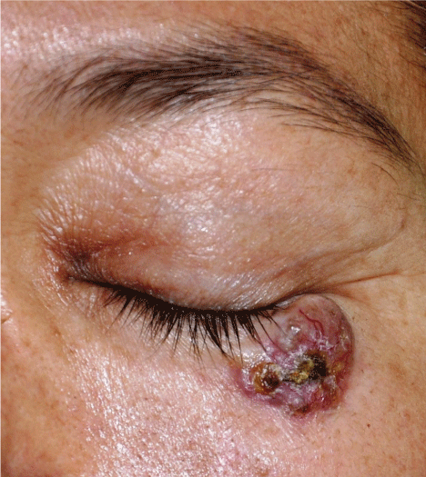
A 51-year-old, female patient, skin phototype IV, presented to the outpatient dermatology clinic for evaluation. She had swelling near the left eye for the past fifteen years with serosanguinous discharge over the last three months. There was gradual increase in the size of the lesion with intermittent pain. Cutaneous examination revealed a 3x2 cm well circumscribed skin tumour with areas of pigmentation, crust and pus discharge. Multiple visible blood vessels and superficial scales were noted on the tumour surface which was firm on palpation.
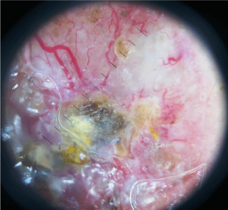
On dermatoscopy atypical red vessels, branching arborizing vessels and milky red background were seen. Histopathology was consistent with Basal Cell Carcinoma (BCC) of the nodulo-cystic variant. She underwent wide local excision and full thickness skin grafting under general anaesthesia (Figure 1).

    Figure 1:  Classical nodular BCC with a central ulcer and surface blood
vessels abutting the lower eyelid.
Arborizing vessels and translucency with a pinkish background have been described as the dermatoscopic hallmarks of nodulocystic BCC. Arborizing telangiectasias is the most characteristic vascular pattern seen in nodular basal cell carcinoma, with a positive predictive value of 94%. Other conditions with arborizing vessels are hidradenoma, intraepidermal poroma and neurothekeoma (Figure 2).
