Special Article - Dermatology Clinical Cases and Images
Austin J Dermatolog. 2015; 2(2): 1039.
Clinical Diagnostic Skill in Dermatology: How to Read Palms: Learning Points from a Routine Clinical Case
Zampetti A1*, Rotoli M2 and Tiberi S3
1Department of Dermatology, Imperial College Healthcare NHS Trust, London, UK
2Department of Dermatology, Università Cattolica del Sacro Cuore,Rome, Italy
3Department of Infectious Diseases, Barts Healthcare NHS Trust, London, UK
*Corresponding author: Anna Zampetti, Imperial College Healthcare NHS Trust, Saint Mary’s Hospital, London, UK
Received: October 16, 2015; Accepted: November 16, 2015; Published: November 18, 2015
Clinical Image
In our outpatient service, a healthy 18-year old girl was seen following the appearance of asymptomatic palm lesions developed in the previous two weeks. The girl did try to apply some creams, including steroids, with no improvement, as also confirmed by the boyfriend who accompanied her. She denied intake of any medications or travel abroad. She was well with no fever or diarrhea and her past medical history was otherwise unremarkable.
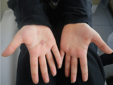
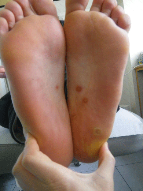
On examination, two erythematous patches were observed in the central area of the right palm. A circular scaly border was noticeable (Figure 1). The face, the trunk and arms were spared and the oral cavity showed no erosion. Permission for pelvic examination was sought and obtained. Annular erythematous asymptomatic plaques were observed on the external genitalia along with some red asymptomatic papular lesions on both soles (Figure 2) which the patient was completely unaware of. Routine laboratory tests such as full blood count, renal and liver function tests, stool culture were ordered; RPR, VDRL, FT-ABS were also requested. These last investigations all confirmed the clinical diagnosis of syphilis. The patient reported that she had her first sexual intercourse with her boyfriend one month before, after many years of engagement and in view of their coming marriage.

    Figure 1:  On the right palm a well defined circular erythematous papular lesion
was noticeable. The lesion shows a peripheral ring of scales, a “collarette”, as
it was first described by Biett. The “Biett’s collarette is a pathognomonic sign
of secondary syphilis.

    Figure 2:  The examination of feet revealed on both soles papular lesions
that along with presence of warty plaques on external genital (condylomata
lata, not shown) supported the clinical impression of syphilis, later confirmed
by serology.
Only a few skin diseases involve palms and soles such as secondary syphilis, Reiter’s disease, psoriasis, drug skin reactions (Steven Johnson’s syndrome, Acute Generalised exanthematous pustolosis- AGEP, Drug Reaction with Eosinophilia and Systemic Symptoms- DRESS), Rocky Mountain spotted fever and Kawasaki syndrome. Secondary syphilis develops 4 to 10 weeks after the appearance of the primary manifestation, an ulcer most commonly on genital area, that can often go unnoticed.
Biett’s collarette, named from the Swiss-born french dermatologist Laurént Thédore Biett who first described it, is a pathognomonic sign of syphilis characterized by a papule encircled by a ring of scales (Figure 1). It can be very helpful to direct investigation especially when facing a diffuse body rash of secondary syphilis, “The Great Imitatator”, or when, as in our case, the skin rash is missing. The communication of the diagnosis was followed by a long discussion about infection transmission and a debate between the girl and his boyfriend. It was clearly explained that syphilis is mainly a sexually transmitted disease, and other routes are rare and unlikely. On the grounds of this information, the partner accepted to be tested for syphilis and although no skin manifestation were previously noted, he was positive to syphilis; HIV, hepatitis B and C serology were negative in both. The couple received benzathine penicillin g 2.4 million units intramuscularly and the regime induced the complete clearing of the skin manifestation on the girl.