Case Report
Austin J Dermatolog. 2016; 3(1): 1043.
Acquired Immunodeficiency Syndrome Patient on Amphetamines with Toxic Epidermal Necrolysis
Yuan-Yu Hou*, Chi-Hsuan Chiang and Feng-Jie Lai
Department of Dermatology, Chi Mei Medical Center, Taiwan
*Corresponding author: Yuan-Yu Hou, Department of Dermatology, Chi Mei Medical Center, Taiwan
Received: December 08, 2015; Accepted: January 15, 2016; Published: January 18, 2016
Abstract
Toxic Epidermal Necrolysis (TEN) is a life threatening disease. Marked keratinocyte apoptosis attributes extensive epidermis detachment. It is mostly drug-related. Aside from the common culprits such as allopurinol, anticonvulsants, antibiotics and Non-Steroid Anti-Inflammatory Drugs (NSAIDs), some reports pointed out that Human Immunodeficiency Virus (HIV) positive patients and amphetamine abusers suffer greater incidence of TEN. We report such a case. Although the pathophysiology of TEN is still veiled, it is believed that complex immune responses play an important role. Further studies for understanding this lethal disease need to be proceeded. Sepsis and multiple organ failure are the major causes of death. Prompt diagnosis and intensive care are imperative.
Keywords:Amphetamine; HIV; AIDS; TEN
Here we report a Human Immunodeficiency Virus (HIV) positive patient on amphetamines with toxic epidermal necrolysis.
Case Presentation
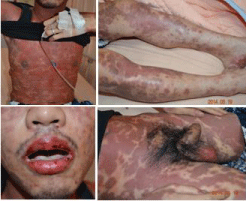
A 24 year-old man who was diagnosed with HIV infection and never received therapy for AIDS. Fever, malaise, sore throat and conjunctivitis proceeded 3 days before skin lesions developed. Patient visited our emergency room, was admitted to burn center thereafter. Generalized painful erythematous to brownish patches with multiple eroded bullae and macules over trunk and extremities were noticed. The skin lesions started from upper trunk and face, and then spread to other parts of the body and extremities, with more than 80% of total Body Surface Area (BSA) involved. Lesions were mostly in erythematous at the beginning, turned darker later on, and started peeling off as it progressed. Blisters/bullae were either flaccid or ruptured. The lesion extended by giving pressure, thus Nikolsky’s sign was positive on physical examination. In addition, oral ulcers, scrotal ulcers were also prominent (Figure 1). Patient denied any medication intake prior to the skin rashes, except amphetamine inhalation about 1 to 2 weeks previous to the above dermatologic examination. Similar but milder episode happened 2 months ago. During the admission period, significant laboratory data : WBC: 2500 /dl (3400-9100 /dL), Hematocrit (Hct): 36.6 % (40-49%), albumin: 3.1 g/dL (3.8-5.3 g/dL), CRP: 15.68 mg/L (< 3 mg/L), hemoglobin : 12.0 g/dL (13.5-17.5 g/ dL), S-GOT(AST)/S-GPT(ALT): 73/65 IU/L (10-50 IU/L), CD4+ lymphocyte counts: 207 /uL (404-1612 /uL), atypical lymphocytes: 2% (=0), CD4+/CD8 ratio: 0.6 (1.2-2.0), HIV viral load test: 125901 copies/ml (non-detected). The vital sign was stable. After admission, no sign of pulmonary embolism/edema, Acute Respiratory Distress Syndrome (ARDS), gastrointestinal hemorrhage, prerenal azotemia, acute tubular necrosis, hypovolemic shock or sepsis appeared.

    Figure 1:  Wide spread maculopapules and bullae, of 80% BSA, with oral and
genital involvement.
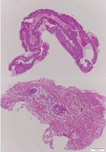
To differentiate the diagnosis of toxic epidermal necrolysis from Generalized Bullous Fixed Drug Eruption (GBFDE), skin biopsy was performed on the lower abdominal area. Pathological findings included: 1. Full-thickness epidermal necrosis with sub epidermal bullae. 2. Sparse inflammatory cells were noted in the dermis (Figure 2), DIF was negative. This above was consistent with the diagnosis of TEN. This patient had been admitted 17 days and given supportive treatment. Patient recovered well, discharged without noticeable sequela except some overt post inflammatory hyperpigmentation residues.

    Figure 2:  Full-layer epidermal necrosis, with sub epidermal bullae formation
and sparse inflammatory cells in dermis.
Discussion
Early differential diagnosis is helpful for making the management strategies and predicting the mortality rate. With severe adverse drug reactions like TEN, that might need intensive care and monitoring. Adequate diagnostic tests and skin biopsy is important.
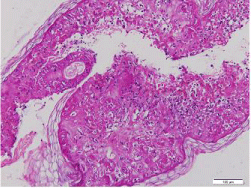
Unlike Stevens - Johnson syndrome (SJS)/TEN, GBFDE is prone to occur in older population. It has shorter latent period. The skin always appears well demarcated erythematous-to-dusky red patches, with less mucosal involvement. Histopathologically, GBFDE shows more melanophages, eosinophils or neutrophils. The infiltration in GBFDE is more superficial and deep rather than superficial infiltration in SJS/TEN. The appearance of aggregated dyskeratotic keratinocyte [1] (Figure 3), so-called fire flag sign in pathology, and Nikolsky’s sign in clinical, both support the diagnosis of TEN.

    Figure 3:  Aggregated dyskeratotic keratinocyte and basal vacuolization in
the epidermis, those are Characteristics of TEN, not GBFDE.
The prevalence of TEN ranges from 0.5-3 person /year/million people, varies from different countries, regions or races, and could have been underestimated [2]. There do exist higher incidence for HIV patients [3-5] or amphetamine abuser [6]. The detailed mechanism remains unclear. In this case, laboratory data showed lowered CD4 helper T cells as well as CD4/CD8 ratio, which is also commonly seen in HIV infected patients. According to Chao Yang, et al. [5], the depleting of CD4 helper T cells predisposes the occurrence of TEN. On the other hand, amphetamine may also play the role to induce TEN in the previous report [6]. Both circumstances could lead to TEN, we are not certain about the exact cause for this patient.
Although prophylactic checking HLA-B1502 for carbamazepine, and HLA-B5801 for allopurinol in preventing SJS/TEN are possible and have actually been performed in some certain countries. It is just for screening of the two culprits. The positive rates differ from races/ regions.
The detection of Fas-s FasL, perforin or granzyme B in SJS/TEN patient is considered to be non-specific. Besides, those procedures need more techniques and facilities. A newer promising measurement of granulysin- a cytotoxic mediator is now available [7]. With quantitative PCR or Immunohistochemistry kits, that can be a better diagnostic tool and predicting factor for TEN.
References
- Cheng-Han Lee, Yi-Chun Chen, Yung-Tsu Cho, Chia-Ying Chang, Chia-Yu Chu. Fixed-drug eruption: A retrospective study in a single referral center in northern Taiwan. Dermatologica Sinica. 2012; 30: 11-15.
- Mittmann N, Knowles SR, Gomez M. Evaluation of the extent of under-reporting of serious adverse drug reactions: the case of toxic epidermal necrolysis. Drug Saf. 2004; 27: 477-487.
- Rzany B, Mockenhaupt M, Stocker U, Hamouda O, Schopf E. Incidence of Stevens-Johnson syndrome and toxic epidermal necrolysis in patients with AIDS in Germany. Arch Dermatol. 1993; 129: 1059-1063.
- Schulz JT, Sheridan RL, Ryan CM, MacKool B, Tompkins RG. A 10-year experience with toxic epidermal necrolysis. J Burn Care Rehabil. 2000; 21: 199-204.
- Chao Yang, Anisa Mosam, Avumile Mankahla, Ncoza Dlova, Arturo Saavedra. HIV infection predisposes skin to toxic epidermal necrolysis via depletion of skin directed CD4+ T cells. Journal of the American Academy of Dermatology. 2014; 70: 1096-1102.
- Hugh Roberts, Alexander Chanberlain, Gordon Rennick, Catriona McLean, Douglas Gin. Severe toxic epidermal necrolysis precipitated by amphetamine use. Australasian Journal of Dermatology. 2006; 47: 114-115.
- Wen-Hung Chung, Shuen-Iu Hung, Jui-Yung Yang, Shih-Chi Su, Shien-Ping Huang, Chun-Yu Wei, et al. Granulysin is a key mediator for disseminated keratinocyte death in Stevens-Johnson syndrome and toxic epidermal necrolysis. Nature Medicine. 2008; 14: 1343-1350.