Clinical Image
Austin J Dermatolog. 2016; 3(2): 1049.
Cutaneous Malignant Fibrous Histiocytoma
Noriega LF1*, Ventura A2 and Pereira GAAM3
Department of Dermatology, Hospital do Servidor Público Municipal de S&aTilde;o Paulo, Brazil
*Corresponding author: Noriega LF, Department of Dermatology, Hospital do Servidor Público Municipal de S&aTilde;o Paulo, 60 Castro Alves, S&aTilde;o Paulo - SP, 01532000. Brazil
Received: March 22, 2016; Accepted: March 29, 2016; Published: March 30, 2016
Keywords
Malignant fibrous histiocytoma; Sarcoma; Skin
Clinical Image
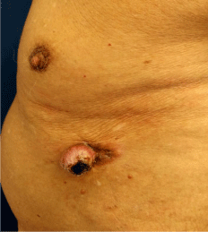
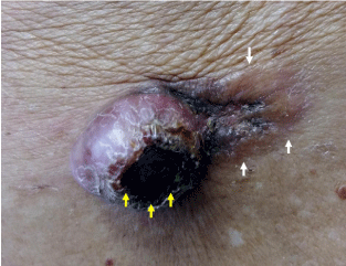
A 93-year-old man presented with an asymptomatic lesion in the left hypochondriac region since 2 years, which showed progressive growth. An erythematous-brownish nodule was observed, measuring 4 cm at its largest diameter, with a superficial haematic crust and perilesional skin infiltration (Figure 1 and 2). The histopathological analysis confirmed the diagnosis of malignant fibrous histiocytoma. En block excision was performed with margins of 2 cm, which resulted in tumor-free surgical margins. The following laboratory and imaging screening were normal. Malignant fibrous histiocytoma is an aggressive subtype of soft tissue sarcoma characterized by a higher incidence in older men. The head and neck, extremities, thorax, and the retroperitoneal space are the most frequently affected. Cutaneous involvement is rare and can be classified into either primary tumour or metastatic tumour, the latter having worse prognosis. In this case, the condition was diagnosed as a superficial primary tumour [1,2].

Figure 1: Nodular lesion in the left hypochondriac region.

    Figure 2:  An erythematous-brownish nodule with a superficial haematic crust
(yellow arrows) and perilesional skin infiltration (white arrows).
References
- Suzuki S, Watanabe S, Kato H, Inagaki H, Hattori H, Morita A. A case of cutaneous malignant fibrous histiocytoma with multiple organ metastases. Kaohsiung J Med Sci. 2013; 29: 111-115.
- Henderson MT, Hollmig ST. Malignant fibrous histiocytoma: changing perceptions and management challenges. J Am Acad Dermatol. 2012; 67: 1335-1341.