Special Article - Dermatology Clinical Cases and Images
Austin J Dermatolog. 2016; 3(2): 1051.
‘Three Zones’ in Reactive Perforating Collagenosis: A New Pattern in Dermoscopy
Ankad BS¹*, Mahajabeen M² and Alekhya R³
Department of Dermatology, S. Nijalingappa Medical College, India
*Corresponding author: Ankad BS, Associate Professor, Department of Dermatology, S. Nijalingappa Medical College, Bagalkot-587103, Karnataka State, India
Received: April 06, 2016; Accepted: April 11, 2016; Published: April 13, 2016
Keywords
Three zones; Dermoscopy; Yellowish-white structureless area; Grayish rim; Blackish halo
Clinical Image
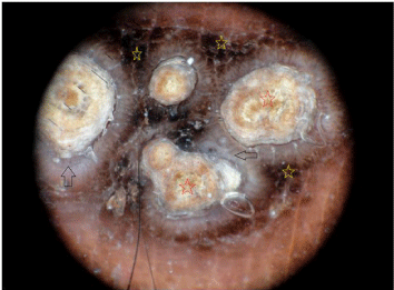
Reactive perforating collagenosis is characterized by umbilicated plaques with crust [1]. Dermoscopic patterns of RPC are described as yellowish-white structureless area in the centre surrounded by grayish rim [2]. Dermoscopy is a new diagnostic tool which allows visualization of surface as well as subsurface structures and it can be used in the diagnosis of acquired perforating dermatoses [3]. Here, new dermoscopic patterns of RPC are described. A 55 year old man, presented with multiple, discrete small, well defined, pruritic ulcerated papules & plaques with yellowish crust for a period of 8 years on the upper and lower extremities as well as posterior trunk (Figure 1). Differential diagnosis included perforating granuloma annulare, prurigo nodularis and other acquired reactive perforating dermatoses. Dermoscopy revealed ‘three zones’ pattern consisting of yellowish-white structureless area in the centre surrounded by grayish rim which is in turn surrounded by blackish halo at the periphery (Figures 2,3). The yellowish-white structureless areas correlate histologically to hyperkeratosis, epidermal hyperplasia, and keratotic plug consisting of collagen, and inflammatory debris (Figure 4). Grayish rim corresponds to epidermal invagination. These patterns are described in earlier study [2]. In this study, authors observed blackish halo surrounding grayish rim. Probably, it represents melanocytes in the lower layers of epidermis which is situated at the edge of crater during the process of invagination. This pattern is not described in the literature. Hence authors propose that ‘three zone’ pattern as a new dermoscopic pattern in reactive perforating collagenosis.

    Figure 1:  Multiple umbilicated papules and plaques with white-yellow crust
affecting lower legs.

    Figure 2:  Dermoscopy demonstrates yellowish-white structureless area (red
stars), grayish rim (black arrows) and peripheral blackish halo (yellow stars).

    Figure 3:  Close up view showing demonstrates yellowish-white structureless
area, grayish rim (yellow arrows) and peripheral blackish halo (yellow stars).

    Figure 4:  Histopathology showing prominent epidermal hyperplasia with
collagen, epidermal invagination and collagen bands (yellow arrow) are
perforating into the epidermis. (10x, H&E).
References
- Kim SW, Kim MS, Lee JH, Son SJ, Park KY, Li K, et al. A clinicopathologic study of thirty cases of acquired perforating dermatosis in Korea. Ann Dermatol. 2014; 26: 162-171.
- Kittisak P, Tanaka M. Dermoscopic findings in a case of reactive perforating collagenosis. Dermatol Pract Concept. 2015; 5: 75-77.
- Ramirez-Fort MK, Khan F, Rosendahl CO, Mercer SE, Shim-Chang H, Levitt JO. Acquired perforating dermatosis: a clinical and dermoscopic correlation. Dermatol Online J. 2013; 19: 1895-1898.