Clinical Image
Austin J Dermatolog. 2016; 3(3): 1052.
Early Detection of Ipilimumab-Induced Hypophysitis using FDG18-PET/CT
Varey AHR and Thompson JF*
Melanoma Institute Australia, The University of Sydney, Australia
*Corresponding author: Thompson JF, Melanoma Institute Australia, The University of Sydney, 40 Rocklands Road, North Sydney NSW 2060, Australia
Received: April 28, 2016; Accepted: May 06, 2016; Published: May 09, 2016
Clinical Image
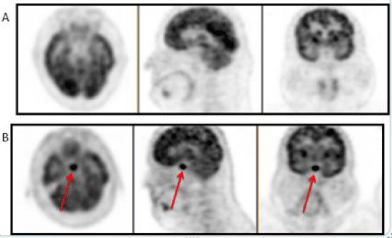
A 46 year old white male with metastatic melanoma was treated with ipilimumab. Following the 4th cycle of treatment, a routine whole body FDG18-PET/CT scan showed strong FDG18 avidity in the pituitary gland, indicative of hypophysitis (panel B) and not present on a pre-treatment scan (panel A) (Figure 1). Although pituitary function was normal at the time of PET/CT, it deteriorated over the next 4 weeks and required treatment with corticosteroids. A brain MRI 4 weeks later confirmed no melanoma metastasis in the pituitary. FDG18-PET/CT scanning may therefore provide early detection of hypophysitis ahead of hormonal derangement, allowing prompt initiation of treatment. The mechanism by which ipilimumab treatment causes hypophysitis is unknown. However, the anterior pituitary, which secretes Adrenocorticotropic Hormone (ACTH) and Melanocyte Stimulating Hormone (MSH), is selectively affected. Both ACTH and MSH are produced by melanoma cells and therefore may be targeted by an ipilimumab-induced immune response.

    Figure 1:  A routine whole body FDG18-PET/CT scan showed strong FDG18
avidity in the pituitary gland, indicative of hypophysitis (panel B) and not
present on a pre-treatment scan (panel A).