Clinical Image
Austin J Dermatolog. 2016; 3(3): 1054.
Dermoscopy in Molluscum Contagiosum: A Novel Diagnostic Aide
Mahajabeen M, Ankad BS*, Alekhya R and Saipriya G
Department of Dermatology, S. Nijalingappa Medical College, India
*Corresponding author: Ankad BS, Associate Professor, Department of Dermatology, S. Nijalingappa Medical College, Bagalkot-587103, Karnataka State, India
Received: June 02, 2016; Accepted: June 09, 2016; Published: June 10, 2016
Keywords
Molluscum contagiosum; Dermoscopy; Telangiectasia; White structureless areas
Clinical Image
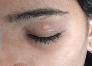
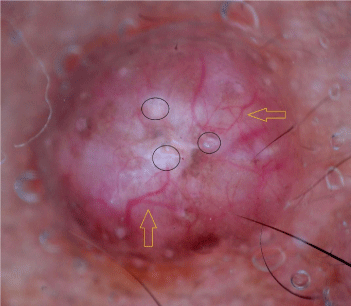
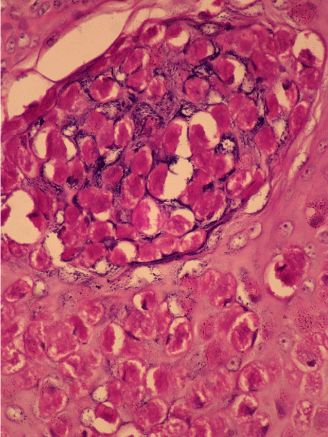
Molluscum Contagiosum (MC) is a viral infection of the epidermal keratinocytes that results in a skin lesion with characteristic intracytoplasmic inclusions caused by pox virus. This common disease is confined to the skin and mucous membranes and produces benign, self limited, multiple umbilicated papular eruptions [1]. Although MC can be diagnosed easily, some factors such as lack of central umbilication, associations with other types of dermatological lesions, atypical lesions, small, single, initial lesions, inflammatory lesions and lesions with perilesional eczema may hamper diagnosis of MC [2]. Therefore, there is a need for early and easy diagnosis of MC. Dermoscopy serves best as a rapid diagnostic aide in such situations. It is a non-invasive diagnostic tool which visualizes subtle clinical patterns of skin lesions and subsurface skin structures that are normally invisible to the naked eyes [3]. A 25 year female presented with asymptomatic, slow growing, skin colored solitary swelling over left upper eyelid since 2 months (Figure 1). There was no history of trauma. Differential diagnosis of sebaceous cyst, inflamed milia, giant MC, nodular basal cell carcinoma and trichilemmoma was made. Dermoscopy showed polylobular whitish structureless areas in the centre surrounded by elongated telangiectasias. Telangiectasias were in serpiginous and arborizing patterns which typically do not cross midline (Figure 2). Excision biopsy was done and histopathology showed pronounced infundibular hyperplasia and papillomatosis with central umbilication. Millions of virions that have proliferated in the cytoplasm of affected epithelial cells result in the characteristic intracytoplasmic bodies that compress the keratinocyte nucleus (Figure 3). The polylobular whitish structureless areas correlate histologically to hyperkeratosis, epidermal hyperplasia, and keratotic molluscum bodies; telangectasia correspond to dilated vessels. Hence, we conclude that dermoscopy plays important role in diagnosing complicated morphology of MC.

Figure 1: Single umbilicated skin colored nodule on the left upper eyelid.

    Figure 2: Dermoscopy shows polylobular white structureless areas (black
circles) in the centre and serpiginous telangiectasias (yellow arrows) at the
periphery.

    Figure 3: Histopathology shows characteristic intracytoplasmic bodies that
compress the keratinocyte nucleus. (H& E, 40x).
References
- Brown J, Janniger CK, Schwartz RA, Silverberg NB. Childhood molluscum contagiosum. Int J Dermatol. 2006; 45: 93-99.
- Ianhez M, Cestari Sda C, Enokihara MY, Seize MB. Dermoscopic patterns of molluscum contagiosum: a study of 211 lesions confirmed by histopathology. An Bras Dermatol. 2011; 86: 74-79.
- Bowling J. Non-melanocytic lesions. Bowling J, editor. In: Diagnostic Dermoscopy - The Illustrated Guide. 1st edn. West Sussex: Wiley-Blackwell. 2012; 59-91.