Case Report
Austin J Dermatolog. 2016; 3(3): 1055.
Vitiligo with Associated Loss of Tattoo Pigments
Oripelaye MM*, Olanrewaju FO, Onayemi O and Olasode OA
Department of Dermatology, Obafemi Awolowo University Teaching Hospitals Complex, Nigeria
*Corresponding author: Oripelaye MM, Department of Dermatology, Obafemi Awolowo University Teaching Hospitals Complex, Ile-Ife, Osun state, Nigeria
Received: April 25, 2016; Accepted: June 22, 2016; Published: June 24, 2016
Abstract
Vitiligo is a common depigmentary skin disorder associated with loss of melanocyte. Since the cause of melanocyte loss is not known, a number of theories such as autoimmune theory, the self-destruct hypothesis and neural hypothesis have been advanced as probable mechanism underlying the melanocyte loss. In this report, we present a ninety three year old woman with observed loss of tattoo pigment on the depigmenting lesions of vitiligo. This observation prompted our suspicion of shared basis for the loss of melanocyte and loss of tattoo pigments.
Keywords: Vitiligo; Tattoo; Melanocyte
Abbreviations
SV: Segmental Vitiligo; NSV: Non-Segmental Vitiligo; MIF: Macrophage Migration Inhibitory Factor
Introduction
Vitiligo is a common acquired depigmentary disorder occurring in 0.4-2.8% of the population [1-3], familial clustering of vitiligo are also commonly reported [4]. It exert a significant negative impact on the quality of life of patients presenting with the disease and even worse in patient with black skin where the contrast is more conspicuous [5].
Various classifications have been described for vitiligo. However, the two main major subtypes are Segmental Vitiligo (SV) and Non- Segmental Vitiligo (NSV). The lesions of vitiligo are characterized by absence of melanocyte. The reason for the loss of melanocyte is not clearly understood however, a number of theories have been postulated to explain the loss of melanocyte although none has been able to explain it satisfactory.
Among the theories postulated to explain the loss of melanocyte are autoimmune theory [6,7], the self-destruct hypothesis [8,9] and neural hypothesis [10]. Other mechanism proposed for loss of melanocyte includes defect in structure and function of endoplasmic reticulum of the melanocyte [11] and primary disturbance of T-lymphocyte [12].
In addition to the above theories, other theories attempting to clarify our understanding of the pathogenesis of vitiligo includes defective melanogenesis with subsequent disappearance of melanocyte, or disappearance of melanocyte due to defective adhesion [13]. The role of Liver X Receptor has been suggested by the finding of increased expression of these receptors on perilesional melanocytes. The increased Liver X Receptor gene expression has been linked to increased susceptibility to vitiligo [14]. The reduced level of antioxidant such as catalase found in patients with vitiligo compared to control depicts the importance of antioxidant in the pathogenesis of vitiligo [15,16].
Since none of the theories standing alone could satisfactorily explain the pathogenesis of vitiligo, Yvon Ganithier et al. have suggested a model integrating all the theories vitiligo [17].
As the cause of vitiligo still remain largely unknown we decided to present a case of vitiligo in which there was loss of tattoo pigment at site of vitiligo lesion.
Case Presentation
We present a ninety three year old African woman who presented to our dermatological clinic with widespread progressively depigmenting skin lesion of ten years duration. The lesion has been increasing progressively from the time it was first noticed ten years ago to its present size involving the shin, arm, forearm, hands, neck and the ear.
There was no associated family history of vitiligo. She is not diabetic, and has no history suggestive of thyroid disease or any other endocrine disease. There is no known precipitant and the patient and family members have been extremely worried by the embarrassment the skin disease has caused them.
She had a cosmetic tattoo about seventy years ago when she was in her twenties and the areas of the skin with the tattoo were the forearm and hands.
The review of other systems was essentially normal. Examination of the skin reveals depigmented patches on the shins, feet, forearms, hands, ear and the chest. Greenish tattoo was also seen on both forearms and dorsum of the hand (Figure 1 & 2).

Figure 1: Tattoo more conspicuous on distal forearm not involved with vitiligo.

    Figure 2: Loss of tattoo symmetry on dorsum of the hands with the left hand
with worse vitiligo affectation having fewer tattoos.
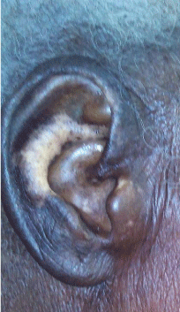
Tattoo in the forearm was observed to be more conspicuous on the area of normal skin, while fading of tattoo was observed in the area of skin involved with depigmentation (Figure 1). On the dorsum of the hands, the tattoo symmetry was lost with right hand being more conspicuous while the left hand with more extensive depigmentation had fewer tattoos (Figure 2). The ear orifices were also involved in the depigmenting process (Figure 3). She also had whitening of hair (probably age rated) (Figure 3), however there is also loss of scalp pigment.

Figure 3: Vitiligo lesion on ear orifice.
Complete blood count was done and was normal range (PCV- 38%, WBC 4,200, neutrophils 65%, Lymphocytes 35%). The blood sugar fasting (5.2mmol/L) and two hour post prandial (5.8mmol/L) were normal.
Assessment of vitiligo was made and the clearing tattoo also noted. The patient and the relatives were counseled on the nature of the diagnosis while phototherapy was being contemplated.
Discussion
Tattoo has been used extensively as camouflage in the management of vitiligo and may continue to be one of the modalities employed in the management of vitiligo. As important as tattoo may look in the management of vitiligo, our interest was aroused by the observation that tattoo was clearing (fading) at the site of the vitiligo. We reasoned that the process involved in the loss of normal skin pigment associated with vitiligo may also be involved in the tattoo clearance. Since tattoo are cause by ingestion of tattoo pigment by macrophages in the dermis, [18] it could be that process that target the destruction of the melanocyte also target the pigment containing macrophages or consume the macrophages in the process.
Serarslan G et al. reported increase macrophage Migration Inhibitory Factor (MIF) in patients with vitiligo [19]. MIF being an activator of macrophage is considered to play significant role in cell mediated immunity. We also reasoned that activated macrophages will have an enhanced potential to clear ingested tattoo pigment. While the role of MIF in vitiligo continues to evolve, it is however established as one of the immunoregulatory cytokines involved in macrophage and T-cell activation and is known to be involved in immune-mediating diseases [20].
Relationship between vitiligo and tattoo was reported by Kluger [21] where he considers a case report of vitiligo on a tattoo as association rather than a cause. Similarly Pan H et al. reported a case of vitiligo induced by tattooing the eyebrow [22]. Our case shows a different relationship where ongoing process of vitiligo was associated with clearance of pre existing tattoo.
The observation of tattoo clearance in our patient may provide newer insight to our understanding of pathogenesis of vitiligo if further researched, and may also influence the role and the methods of tattoo use in the management of vitiligo.
References
- Hann SK, Nordlund JJ. Definition of vitiligo. Hann SK, Nordlund JJ, editors. In: Vitiligo. London, Blackwell Science. 2000; 3.
- Nordlund JJ, Ortonne JP. Vitiligo and depigmentation. WL Weston, editor. In: Current Problems in Dermatology. St Louis, Mosby-Year Book. 1992.
- Nanette BS. The epidemiology of vitiligo. Current dermatology report. 2015; 4: 36-43.
- Karelson M, Silm H, Salum T, Koks S, Kingo K. Differences between familial and sporadic cases of vitiligo. J Eur Acad Dermatol Venereol. 2012; 26: 915-918.
- Linthorst Homan MW, Spuls PI, de Korte J, Bos JD, Sprangers MA, van der Veen JP. The burden of vitiligo: Patients characteristics associated with quality of life. J Am Acad Dermatol. 2009; 61: 411-420.
- Bystryn JC. Theories on the pathogenesis of depigmentation: Immune hypothesis. Hann SK, Nordlund JJ, editors. In: Vitiligo. London, Blackwell Science. 2000; 129.
- Norris DA, Kissinger RM, Naughton GM, Bystryn JC. Evidence for immunologic mechanism in human vitiligo: patientís sera induce damage to human melanocyte in vitro by complement-mediated damage and antibody-dependent cellular cytotoxicity. J Invest Dermatol. 1988; 90: 783-789.
- Schallreuter KU. Biochemical theory of vitiligo: A role of pteridines in pigmentation. Hann SK, Nordlund JJ, editors. In: Vitiligo. London, Blackwell Science. 2000; 151.
- Moellmann G, Klein-Angerer S, Scollay DA, Nordlund JJ, Lerner AB. Extracellular granular material and degeneration of keratinocytes in the normally pigmented epidermis of patients with vitiligo. J Invest Dermatol. 1982; 79: 321-330.
- Orecchia GE. Neural pathogenesis. Hann SK, Nordlund JJ, ediotors. In: Vitiligo. London, Blackwell Science. 2000; 142.
- Boissy RE. The intrinsic (genetic) theory for the cause of vitiligo. Hann SK, Nordlund JJ, editors. In: Vitiligo. London, Blackwell Science, 2000; 123.
- Ortonne JP, Bose SK. Vitiligo: Where do we stand? Pigment Cell Res. 1993; 6: 61-72.
- Morelli JG, Yohn JJ, Zekman T, Norris DA. Melanocyte movement in vitro: role of matrix proteins and integrin receptors. J Invest Dermatol. 1993; 101: 605-608.
- Agarwal S, Kaur G, Randhawa R, Mahajan V, Bansal R, Changotra H. Liver X Receptor-a polymorphisms (rs11039155 and rs2279238) are associated with susceptibility to vitiligo. Meta Gene. 2016; 8: 33-36.
- Schallreuter KU, Wood JM, Berger J. Low catalase levels in the epidermis of patients with vitiligo. J Invest Dermatol. 1991; 97: 1081-1085.
- Casp CB, She JX, McCormack WT. Genetic association of the catalase gene (CAT) with vitiligo susceptibility. Pigment Cell Res. 2002; 15: 62-66.
- Gauthier Y, Cario Andre M, Taieb A. A critical appraisal of vitiligo etiologic theories. Is melanocyte loss a melanocytorrhagy? Pigment Cell Res. 2003; 16: 322-332.
- Anne L. Body Art. Klauss W, Lowell AG, Stephen K, Barbara AG, Amy SP, David JL, editors. In: Fitzpatrick's Dermatology in General Medicine. 7th Edn. New York: McGraw-Hill Professional. 2008; 887.
- Serarslan G, Yonden Z, Sogut S, Savas N, Celik E, Arpaci A. Macrophage migration inhibitory factor in patients with vitiligo and relationship between duration and clinical type of disease. Clin Exp Dermatol. 2010; 35: 487-490.
- Shimizu T. Role of macrophage Migratory Inhibition Factor (MIF) in the skin. J Dermatol Sci. 2008; 49: 95-97.
- Kluger N. Vitiligo on a tattoo: association rather than cause. International Journal of Dermatology. 2013; 52: 1617-1618.
- Pan H, Song W, Xu A. A case of vitiligo induced by tattooing eyebrow. Int J Dermatol. 2011; 50: 607-608.