Case Report
Austin J Dermatolog. 2016; 3(3): 1056.
Tinea Capitis Mimicking as Cicatricial Alopecia of Scalp
Muhammad J, Hafeez ud Din, Ahmed K and Anwar MI*
Department of Dermatology, Bahria University Medical & Dental College, Pakistan
*Corresponding author: Muhammad Irfan Anwar, Department of Dermatology, Bahria University Medical & Dental College, Karachi, Pakistan
Received: June 07, 2016; Accepted: June 27, 2016; Published: June 29, 2016
Abstract
Tinea capitis is a very common childhood fungal infection of hair follicles. It clinically manifests as asymptomatic scaling, inflammatory suppurative nodules, draining sinuses and tracks. Secondary cicatricial alopecia may occur in tinea capitis as a result of chronic persistent infection. Rarely, initial presentation can mimic primary cicatricial alopecia. We herein, report a case of 9 years old healthy Asian female with chronic tinea capitis masquerading as cicatricial alopecia. Prompt diagnosis and management is imperative to prevent permanent scarring alopecia.
Keywords: Tinea capitis; Cicatricial alopecia; Hair follicles; Scalp
Introduction
Tinea capitis is the most common dermatophyte infection in children. Trichophyton and Microsporum species are common causative organisms worldwide. Clinical spectrum varies from asymptomatic carriage to symptomatic and inflammatory disease. It can mimick with seborrheic dermatitis of scalp and can at times become difficult to diagnose. Black dot type of tinea captitis is mostly caused by trichophyton species and they usually present as annular scaly erythematous patches having broken hairs. Draining sinus tracts, thick crusts, boggy plaques and suppurative nodules are seen in Favus and Kerion type of tinea capitis. Oral antifungal agents are the mainstay of therapy in all types of tinea capitis, Owing to their ability to penetrate the hair follicle and shaft. If left untreated or in cases of chronic severe inflammation, cicatricial alopecia ensues serious complication because hair follicles are permanently destroyed. Uncommonly, an initial presentation can mimic primary cicatricial alopecia [1]. To our knowledge this is the first case report of tinea capitis presenting as primary cicatricial alopecia from Pakistan.
Case Presentation
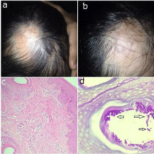
A 9 year old Pakistani female with no known co morbidities, presented with a 06 years history of patchy hair loss from scalp. Detailed history revealed that initially she developed erythema, mild scaling and pruritis of scalp along with slowly progressive loss of hair, which ultimately resulted in permanent hair loss. She had no constitutional symptoms and no family member affected. However there was a history of contact with healthy cats at home. There was no significant drug history. She was neglected by her parents regarding proper treatment of her disease due to lower socio-economic background and had only been using anti dandruff shampoos with no improvement in her condition. Her scalp examination revealed a scarred patch of alopecia without erythema and mild scaling at the periphery of patch (Figure1- a & b). Hair pull test was negative. Wood’s lamp examination of scalp did not reveal any fluorescence. KOH preparation of scales was negative for fungal hyphae. There was no oral, mucosal and nail changes and regional lymph nodes examination was unremarkable.

    Figure 1:  a & b: Patches of scarring alopecia over scalp, c: Histopathology
of scarring alopecia showing perifollicular lympocytic infiltrate with dense
papillary dermis fibrosis, d: PAS staining showed magenta colored spores
and hyphae in hair follicle (marked with black arrows).
A scalp biopsy was done and tissue examined revealed unremarkable epidermis, loss of hair follicles and perifollicular lymphocytic infiltrate with dermal fibrosis (Figure1- c). PAS stain showed fungal spores and hyphae in hair follicles (Figure1- d) thus confirming the diagnosis of Tinea capitis. Unfortunately, fungal culture facility was not available in laboratory and griseofulvin was not available in local pharmacies; so her treatment was started with oral terbinafine (125 mg/day) and topical terbinafine application overnight, with morning wash with 02% ketoconazole shampoo, to which she responded very well. No more hair loss and expansion of alopecia patches were noted on follow up examination. The scaling at the margins of patches are also absent on follow up exam. Unfortunately, no more hair regrowths was seen in scarred patches.
Discussion
Tinea capitis is the most common dermatophyte scalp infection in children [1]. The most common organism involved in endemic cases is “Trichopyton tonsurans” [2]. Endothrix caused by Trichophyton tonsurans invade inside of hair shaft. Ectothrix fungi invade outside of hair shaft, being caused by various Microsporum species which characteristically are fluorescent. Favus infection is caused by Trichophyton schoenlenii having hyphae which invade hair shaft and produce linear air spaces [3]. Clinically, various patterns of tinea capitis are seen. Seborrheic pattern presents with dandruff like scaling and should be considered in pre-pubertal children having seborrheic dermatitis type presentation. Presence of black dots in bald area and hair breakage at scalp line characterizes “Black dot” type tinea capitis. Kerion manifests as tender boggy mass and posterior cervical lymphadenopathy mimicking bacterial folliculitis and abscess. Cup shaped honey colored crust which are called scutula and alopetic areas are seen in rare inflammatory type of tinea capitis called “Favus” [4].
The amount of scale may be significant or there may be no scaling, and its color may be white, yellow or brown. Hair loss is minimal or absent or it may be remarkable with scarring. Broken hairs that resemble “black dots” may or may not be present, and can be scattered among normal hairs or concealed by scale. The inflammatory type of tinea capitis presents with marked edema, redness, pustules, nodules, or sinus tracts with purulent discharge and crusting. Hair loss may be patchy or involve the entire scalp. Inflammatory lesions may be tender with marked cervical lymphadenopathy and can be associated with systemic symptoms. Diagnosis is confirmed by wood’s lamp fluorescence, microscopic detection of fungal elements using KOH preparation of hair shaft, histopathologic evidence of hyphae in hair follicles using PAS stain or species identification by fungal culture [5].
Even though KOH examination can provide a rapid office diagnosis of tinea capitis. This case highlights the fact that a negative KOH preparation does not exclude a diagnosis of tinea capitis. Although fungal culture is a vital part of the evaluation of chronic hair loss in childhood but lack of fungal culture facility was a real time issue in our case. Although oral griseofulvin is the gold standard treatment for tinea capitis, treatment failures are commonly reported because of resistance and intolerant side effects. However, in our case we could not use it because of local unavailability of drug in market. Newer antifungal agents like Itraconazole and terbinafine are similar to griseofulvin in efficacy and are safe in pediatric population. Kerion in addition to oral antifungal are treated with wet dressings to remove crust, analgesia and antibiotics for coexist ant bacterial infections [6]. Adjunctive treatment with topical 02% ketoconazole shampoo/ and 01% selenium sulfide shampoos has been suggested to reduce the shedding of fungal spores and is recommended for patients. Topical terbinafine may be used as adjunctive therapy if the fungal burden is thought to be high. Family members of patient should undergo evaluation for infection or carrier state [4]. Carriers are treated with antifungal shampoos because if these left untreated will decrease cure rates for tinea capitis [7]. Treating domestic pets with griseofulvin is also recommended [8]. All her family members underwent clinical examination but none had signs related to disease. We have counseled her parents regarding strict adherence to treatment, follow up and to improve standards of pet hygiene.
This case report highlights the importance of an early recognition of these common fungal infections in children to prevent permanent disfiguring complications like scarring alopecia.
References
- Mirmirani P, Willey A, Chamlin S, Frieden IJ, Price VH. Tinea Capitis mimicking Cicatricial alopecia: What host and dermatophyte factors lead to this unusual presentation? J Am Acad Dermatol. 2009; 60: 490-495.
- Foster KW, Ghannoum MA, Elewski BE. Epidemiologic surveillance of cutaneous fungal infections in the United States from 1999 to 2002. J Am Acad Dermatol. 2004; 50: 748-752.
- Fitzpatrick J, Morelli J. Dermatology Secrets Plus. 4th Edn: Elsevier Mosby. 2011; 31: 217-218.
- Sobera JO, Elewski BE. Fungal. Bolognia JL, Jorizzo JL, Rapini RP, editors. In: Fungal Infections. Dermatology, New York. 2008; 1135.
- Goldsmith LA, Katz S, Gilchrest B, Paller A, Leffell D, Wolff K. Fitzpatrick’s Dermatology in General Medicine. 8th Edn: MC Graw Hill. 2012; 2: 2280.
- Griffiths C, Barker J, Bleiker T, Chalmers R, Creamer D. Rook’s Textbook of Dermatology. 9th Edn: Blackwell Wiley. 2016; 32: 9-41.
- Babel D, Baughman S. Evaluation of the adult carrier state in juvenile tinea capitis caused by Trichophyton Tonsurans. J Am Acad Dermatol. 1989; 21: 1209-1212.
- Neil G, Hanslo D, Buccimazza S, Kibel M. Control of the carrier state of scalp dermatophytes. Pediatr Infect Dis J. 1990; 9: 57-58.