
Case Report
Austin J Dermatolog. 2016; 3(4): 1058.
Blue Rubber Bleb Nevus Syndrome Manifest as Chronic Anemia in a 17-Year-Old Boy
Zhong M¹, Song Z²*, Chen W³, Yang X² and Hao F²
¹Department of Dermatology, Outpatient Department, People’s Hospital, China
²Department of Dermatology, Third Military Medical University, China
³Department of Dermatology and Allergy, Technische Universitaet Muenchen, Germany
*Corresponding author: Zhiqiang Song, Department of Dermatology, Southwest Hospital, Third Military Medical University, Chongqing 400038, China
Received: August 16, 2016; Accepted: October 03, 2016; Published: October 05, 2016
Abstract
Blue Rubber Bleb Nevus Syndrome (BRBNS) is a rare systemic disorder characterized by cutaneous and Gastrointestinal (GI) vascular malformations that sometimes lead to occult blood loss with iron-deficiency anaemia or severe gastrointestinal bleeding. Here we report a case of a 17-year-old patient with BRBNS manifest as chronic anemia in his early years and review the clinical features and treatment of this rare disorder.
Keywords: Blue rubber bleb nevus syndrome; Bean syndrome; Anemia; Endoscopy
Case Report
A 17-year-old boy was admitted to department of pediatrics with ten years history of dizzy, lassitude, inertia. His chief complaint was dizziness that had initially developed 10 years ago. The patient did not pay more attention to this because it did not affect his study and life. Occasionally he felt cardiopalmus, upper abdominal pain but without fever, hematemesis and melena. After the illness, the patient had slightly poor memory and appetite and decreased body weight. Five years ago, his parent found a few blue eruptions on his trunk. Although more lesions appear with time, they did not take the patient to see a dermatologist, because they are told these lesions were nevi and did not need further treatments.
Physical examination revealed the patient presented with multiple violaceous or blue cutaneous lesions with various diameter on the face, trunk and the palmar surface of both feet. The lesions were compressible; after compression they slowly refilled with blood (Figure 1).
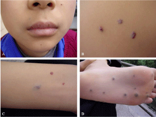
    Figure 1:  Multiple bluish-black lesions on the face (A), trunk (B), arm (C)
and foot (D).
Hemoglobin level at admission was 33.2g/L (normal: 110-160 g/L). Red blood cell (RBC) count was 2.28×10¹²/L (normal: 3.5-5.5×10¹²/L). Mean Corpuscular Volume (MCV), Mean Corpuscular Hemoglobin (MCH) and Mean Corpuscular Hemoglobin Concentration (MCHC) was 60.5fl (normal: 80~100fL), 14.5pg (normal: 26-38pg), and 239 g/L (normal: 310-370 g/L), respectively. White blood count and platelet count were within normal limits. Liver function test, renal function test, routine urine and sedes examination were all normal.
After a consultation by dermatologist, the patient was given the diagnosis with blue rubber bleb nevus syndrome. He then received upper endoscopy and biopsy of skin eruption from his trunk. Upper endoscopy disclosed multiple purplish and compressible polypoid lesions on the esophagus and stomach with 0.5-1cm in diameter. Actively oozing blood was not seen (Figure 2). Histological examination revealed multiple dilated venous channels filled with blood and lined by flattened endothelium and an attenuated smooth muscle coat in dermis and subcutis (Figure 3).
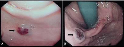
    Figure 2:  Endoscopic views showed polypoid lesions on the esophagus and
stomach (A and B).
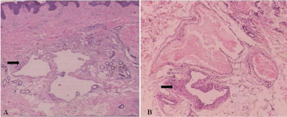
    Figure 3:  Multiple dilated venous channels filled with blood in dermis and
subcutis. (Hematoxylin-eosin stain, A×40, B×100).
The patient received system therapy including blood transfusion, oral ferrous sulfate and vitamin B12. Three weeks later, his symptom with dizzy, lassitude, inertia improved gradually. Hemoglobin level and RBC count returned to 75.2g/L and 3.58×10¹²/L, respectively. Treatment continued after he left hospital. Half one year later, the symptoms with dizzy, lassitude, inertia improved completely, and hemoglobin level and RBC count return to normal limits. Signs of bleeding complications were not found by endoscopic examination. Long term follow-up is in progress.
Discussion
The majority of Blue Rubber Bleb Nevus Syndrome (BRBNS) is sporadic, but it may be passed on as an autosomal dominant condition and associated with the TEK tyrosine kinase receptor which involved in endothelial-smooth muscle cell signaling [1]. Symptoms and signs may be different according to the organ involved, but the most common clinical manifestation is iron deficiency anemia secondary to overt bleeding episodes, especially when this disease is under diagnosis.
Cutaneous lesions of BRBNS are usually present at birth or in childhood, whereas GI involvement develops in adulthood [1-3]. However, this does not mean absolute uniform in order. As seen in our case, GI involvement may develop earlier than cutaneous lesions, and thus be correlated with unnoticed chronic anemia. The cutaneous lesions are found most commonly on the trunk and upper extremities, which may be macular, papular, nodular and characterized by their small size, bluish color, softness and the tendency to refill with blood after compression. The lesions are usually asymptomatic and rarely bleed spontaneously but are easily traumatized, in contrast to the GI lesions, which sometimes bleed spontaneously.
GI lesions are most commonly found in the small intestine and distal large bowel and presented as discrete bluish mucosal nodules [4]. Before skin lesions occur, patient may present with anemia caused by frank or occult bleeding from vascular lesions in the GI tract, and would lead the patient be admitted to department of internal medicine or pediatrics, as in our case. Sometimes, the earlier cutaneous eruptions are small and less, and such condition is commonly ignored by internist and/or pediatrician, and would lead to the misdiagnosis and the neglect of the underlying cause of anemia.
The diagnosis of BRBNS was made by considering the clinical and histological findings. In rare instances, BRBNS can involve other organs such as lungs, spleen, liver, kidneys, bladder, brain and skeletal muscle [1,4]. Radiographic images and Magnetic Resonance Imaging (MRI) are useful for screening possible extracutaneous lesions.
Treatment for BRBNS is usually based on the extent of GI involvement and the severity of the clinical symptoms, especially subsequent anaemia [2,3]. It is reported that the number of skin and GI lesions and the severity of anaemia may correlated [1,2]. If the patient has no obvious signs of massive bleeding, conservative management with iron supplementation or blood transfusion will be sufficient [5]. If active and or massive bleeding occurs, individual surgical excision of the gut lesions has been demonstrated to have long-term efficacy in eliminating bleeding. Segmental resection of involved gut has been debated because this would lead to both short intestine and continued bleeding from the unresected lesions [1,6].
We want to present this case particularly to emphasize the possibility of chronic anemia in BRBNS and heighten physicians awareness about the disease, and to show a conservative treatment could get enough and long term clinical effects for cases with less numbers of skin and GI lesions and without obvious anaemia.
Acknowledgement
This work was supported in part by Clinical Research Funding of Southwest Hospital, SWH2013LC10 and Natural Science Foundation Project of Chongqing, cstc2015jcyjA10118.
References
- Feingold RM. The blue rubber bleb [corrected] nevus syndrome. J Insur Med. 2009; 41: 67-71.
- Massoumi H, Patel S. Blue rubber bleb nevus syndrome. Gastrointestinal Endoscopy. 2007; 65: 1076-1077.
- Ertem D, Acar Y, Kotiloglu E, Yucelten D, Pehlivanoglu E. Blue rubber bleb nevus syndrome. Pediatrics. 2001; 107: 418-420.
- Mechri M, Soyer P, Boudiaf M, Duchat F, Hamzi L, Rymer R. Small bowel involvement in blue rubber bleb nevus syndrome: MR imaging features. Abdom Imaging. 2009; 34: 448-451.
- Boente MD, Cordisco MR, Frontini MD, Asial RA. Blue rubber bleb nevus (Bean syndrome): evolution of four cases and clinical response to pharmacologic agents. Pediatr Dermatol. 1999; 16: 222-227.
- Domini M, Aquino A, Fakhro A, Tursini S, Marino N, Di Matteo S, et al. Blue rubber bleb nevus syndrome and gastrointestinal haemorrhage: which treatment? Eur J Pediatr Surg. 2002; 12: 129-133.