Clinical Image
Austin J Dermatolog.2016; 3(4): 1061.
Dermoscopy Picks Porokeratosis: A Case Report
Ankad BS¹* and Savitha LB²
Department of Dermatology, S. Nijalingappa Medical College, India
*Corresponding author: Ankad BS, Department of Dermatology, S. Nijalingappa Medical College, Bagalkot-587103, Karnataka State, India
Received: November 14, 2016; Accepted: November 21, 2016; Published: November 22, 2016
Keywords
Dermoscopy; Porokeratosis; Patterns
Clinical Image
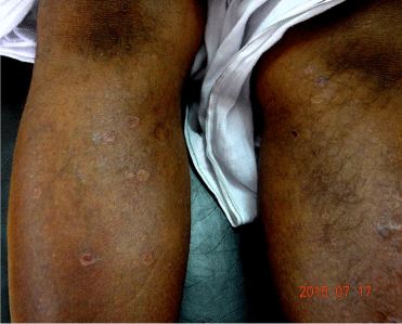
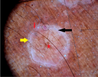
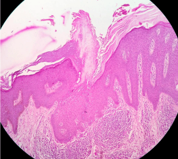
Porokeratosis are a group of disorders of keratinization characterized by annular lesions surrounded by a characteristic keratotic border which corresponds to a typical histopathologic feature, namely, the coronoid lamella [1]. Dermoscopy visualizes subsurface structures in a magnified view with certain characteristic color patterns which helps in the clinical diagnosis [2]. Here authors describe a case scenario wherein dermoscopy aided the diagnosis of superficial actinic porokeratosis. A 45 year male presented with multiple, round to oval shaped skin lesions over both hands and legs with mild itching observed in last 15 days. Examination revealed erythematous to brownish circular plaques with raised borders with central atrophy distributed symmetrically over extensors of both hands and legs (Figure 1). Clinically, superficial actinic porokeratosis, annular lichen planus, disseminated granuloma annulare and lichen planus atrophicus were suspected. Dermoscopy was performed on the lesion over the leg using DermLite 3 Gen with polarized lights and 10x magnifications. It demonstrated white track structure with central brown single (sometimes double) line at the periphery. Perifollicular whitish areas were observed in the periphery. Red globules were present at periphery and brown areas were observed at the centre (Figure 2). Histopathology of the lesion revealed characteristic coronoid lamella with thin column of parakeratotic cells with an absent or decreased underlying granular zone (Figure 3). Based on clinical, dermoscopic & histopathological evidence diagnosis of superficial actinic porokeratosis was made. White track structure and central brown line corresponds to coronoid lamella and acanthotic epithelium. Red globules correspond to dilated capillaries below the atrophic epidermis and brown areas represent melanin in histopathology [3]. Other dermoscopic patterns of porokeratosis are pink-white scar-like area in the center [2] and white homogenous area [3]. However, these patterns were not observed in this case. Dermoscopy of annular lichen planus and lichen planus atrophicus shows pearly white areas in starbursts pattern, diffusely arranged blue-gray globules and peripheral red globules [4]. Granuloma annulare demonstrates reddish yellow color at periphery surrounding a structureless area [5]. Although above mentioned conditions were considered as differentials in the clinical diagnosis, dermoscopy could pick the correct diagnosis by showing characteristic patterns of superficial actinic porokeratosis. Hence, dermoscopy being a noninvasive technique can be a quick office procedure in distinguishing porokeratosis from other annular lesions in clinical practice.

    Figure 1:  Clinical image showing annular atrophic plaques on the anterior
leg.

    Figure 2:  Dermoscopy showing brown line (yellow arrow) surrounded by
white tract structure (black arrow) at periphery. Perifollicular whitish areas
(red arrow) and red globules (black circle) are seen in the periphery. Brown
areas (red star) are present in the centre. [10x magnification, polarized mode].

    Figure 3:  Histopathology shows characteristic hyperkeratosis, parakeratotic
coronoid lamella and acanthosis in the epidermis. (H & E, 10x).
References
- Delfino M, Argenziano G, Nino M. Dermoscopy for the diagnosis of porokeratosis. J Eur Acad Dermatol Venereol. 2004; 18: 194-195.
- D’Amico D, Vaccaro M, Guarneri C, Borgia F, Cannavó SP, Guarneri F. Video dermatoscopic approach to porokeratosis of Mibelli: a useful tool for the diagnosis. Acta Derm Venereol. 2001; 81: 431-432.
- Zaballos P, Puig S, Malvehy J. Dermoscopy of disseminated superficial actinic porokeratosis. Arch Dermatol. 2004; 140: 1410.
- Lallas A, Kyrgidis A, Tzellos TG, Apalla Z, Karakyriou E, Karatolias A, et al. Accuracy of dermoscopic criteria for the diagnosis of psoriasis, dermatitis, lichen planus and pityriasis rosea. Br J Dermatol. 2012; 166: 1198-1205.
- Lallas A, Zaballos P, Zalaudek I, Apalla Z, Gourhant JY, Longo C, et al. Dermoscopic patterns of granuloma annulare and necrobiosis lipoidica. Clin Exp Dermatol. 2013; 38: 425-427.