Case Report
Austin J Dermatolog. 2017; 4(2): 1075.
Rare Location of a Trichoblastic Carcinoma: A Case Report
Lahbali O*, Azami MA, Tbouda M, Bourhoum N, Znati K and Mahassini N
Department of Pathology, University Hospital Ibn Sina, Morocco
*Corresponding author: Othmane Lahbali, Department of Pathology, University Hospital Ibn Sina, Mohammed V University in Rabat, Morocco
Received: June 12, 2017; Accepted: July 03, 2017; Published: July 11, 2017
Abstract
Trichoblastic carcinoma is a rare malignant adnexal tumor that can give metastases. It occurs mainly in the elderly and often poses diagnostic problems with basal cell carcinoma. We report the observation of a 50-year-old man who consulted for an ulcerated lesion of the abdomen evolving for 1 year. A surgical excision was performed and the histological analysis showed a trichoblastic carcinoma associated with a trichoblastoma. The particularity of our case is the entity which is rare and its location exceptional, since only one case has been reported in the abdominal level in the literature.
Keywords: Trichoblasctic carcinoma; Adnexal tumor; Basal cell carcinoma
Introduction
Trichoblastic carcinoma is an aggressive malignant epithelial tumor of the follicular germ. It may develop on lesions of solitary or multiple trichoblastoma.
This rare entity is characterized by a very important recurrent and metastatic power and often poses a differential diagnosis problem with basal cell carcinoma.
Case Presentation
We report a case of a 50-year-old man with an ulcerated lesion in the peri-umbilical abdominal level evolving for 1 year. The clinical examination found an ulcerated indurate lesion measuring 4cm long. Surgical excision was performed with a safety margin of 1cm.
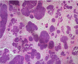
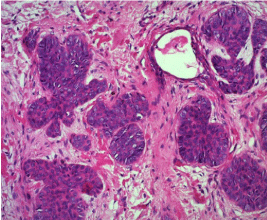
The microscopic examination showed a tumor proliferation composed of a double contingent. The first contingent contains epithelial lobules of small and medium size within a cellular stroma (Figure 1). The tumor cells are more basophilic at the periphery of the epithelial lobules than at the center (Figure 2).

    Figure 1:  Trichoblastoma: the tumor is composed of basophilic lobules
surrounded by a dense fibrous stroma. Keratocysts are not present
(Haematoxylin and eosin stain (HE), magnification x 50).

    Figure 2:  Trichoblastoma: the tumor is composed of small cells with uniform
darkly staining nuclei (Haematoxylin and eosin stain, magnification x 200).
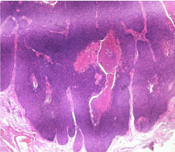
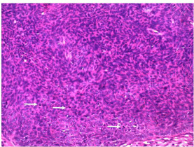
The second contingent is composed of epithelial massifs (Figure 3) which infiltrate the adjacent structures at depths and laterally. The tumor cells that compose it are basophils showing several figures of mitoses (Figure 4). The diagnosis that was retained is that of a trichoblastic carcinoma developed on trichoblastoma. An assessment of extension has been made and which has not objectified secondary localization.

    Figure 3:  Trichoblastic carcinoma: Large infiltrating epithelial lobules
(Haematoxylin and eosin stain, x 50).

    Figure 4:  Trichoblastic carcinoma showing several mitosis figures (arrow)
(Haematoxylin and eosin stain, x 200).
Discussion
Trichoblastic carcinoma is a rare malignant adnexal tumor; about twenty one cases have been reported in the literature [1,2].
Mean age at the time of presentation seems to be lower than basal cell carcinoma. They are located on all the integuments including in non-classical areas for basal cell carcinomas, such as limbs or the trunk [3,4]. The peculiarity of our case is its peri-umbilical abdominal localization only one case has been reported in the literature in this location [5].
Clinically this lesion is often of large size ulcerated and can occur on a pre-existing trichoblastoma [6].
The diagnosis is based on the microscopic examination of the epithelial lobule comprising basaloid cells with a pilar differentiation characterized by central keratinization and a cellular stroma.
Mitosis and necrosis may be present in both the benign and malignant component, so the diagnosis of malignancy is mainly posed by the infiltrating power of adjacent structures with the possibility of tumor vascular emboli.
The differential diagnosis is mainly with basal cell carcinoma. Trichoblastic carcinoma can give nodal and visceral metastases whose prevalence remains unknown [7,8]. In comparison, basal cell carcinoma only metastasizes exceptionally (0.003 to 0.1%) [9]. The development of a trichoblastic carcinoma on a pre-existing trichoblastoma raises the hypothesis of a malignant degeneration of a trichoblastoma [6].
There are no clear recommendations on therapeutic management of trichoblastic carcinoma (surgical safety margins or complementary therapies), due to the low number of cases published.
Conclusion
Trichoblastic carcinoma is a rare malignant adnexal tumor which often poses a differential diagnosis problem with basal cell carcinoma and whose treatment is based on a wide surgical excision with sufficient margins to avoid the risk of local recurrence and distant metastasis.
References
- Leblebici C, Altinel D, Serin M, Okcu O, Yazar SK. Trichoblastic carcinoma of the scalp with rippled pattern. Indian J Pathol Microbiol. 2017; 60: 97-98.
- Triaridis S, Papadopoulos S, Tsitlakidis D, Printza A, Grosshans E, Cribier B. Trichoblastic carcinoma of the pinna. A rare case. Hippokratia. 2007; 11: 89-91.
- Laffay L, Depaepe L, d’Hombres A. Histological features and treatment approach of trichoblastic carcinomas: from a case report to a review of the literature. Tumori. 2012; 98: 46e-49e.
- Ayhan M, Gorgu M, Aytug Z. Trichoblastic carcinoma of the alar region: a case report. Dermatol Surg. 2006; 32: 976-979.
- Sau P, Lupton GP, Graham JH. Trichogerminoma. Report of 14 cases. J Cutan Pathol. 1992; 19: 357-365.
- Schulz T, Proske S, Hartschuh W. High-grade trichoblastic carcinoma arising in trichoblastoma: a rare adnexal neoplasm often showing metastatic spread. Am J Dermatopathol. 2005; 27: 9-16.
- Battistella M, Mateus C, Lassau N. Sunitinib efficacy in the treatment of metastatic skin adnexal carcinomas: report of two patients with hidradenocarcinoma and trichoblastic carcinoma. J Eur Acad Dermatol Venereol. 2010; 24: 199-203.
- Regauer S, Beham-Schmid C, Okcu M, Hartner E, Mannweiler S. Trichoblastic carcinoma (‘‘Malignant Trichoblastoma’’) with lymphatic and hematogenous metastases. Mod Pathol. 2000; 13: 673-678.
- Von Domarus H, Stevens PJ. Metastatic basal cell carcinoma. Report of five cases and review of 170 cases in the literature. J Am Acad Dermatol. 1984; 10: 1043-1060.