
Special Article - Diagnosis and Usefulness of Dermoscopy
Austin J Dermatolog. 2017; 4(2): 1076.
Acral Melanoma Showing Fibrillar Pattern on Dermoscopy
Togawa Y*, Wakabayashi S, Suehiro K and Matsue H
Department of Dermatology, Chiba University of Graduate School of Medicine, Japan
*Corresponding author: Yaei Togawa, Department of Dermatology, Chiba University of Graduate School of Medicine, 1-8-1, Inohana, Chuo-ku, Chiba, 260-8670, Japan
Received: September 11, 2017; Accepted: October 04, 2017; Published: October 11, 2017
Keywords
Acral melanoma; Dermoscopy; Fibrillar pattern
Clinical Image
A 50-year-old female presented with a black macule on the heel that appeared 2 years ago. Clinical examination revealed a black macule with ill-defined border on the lateral side of the sole, measuring 13 x 10mm. Dermoscopic examination showed Fibrillar Pattern (FP) with irregular black dots in the center and a parallel ridge pattern in the periphery (Figure 1). The lesion was removed with a 5mm margin and pathologically diagnosed as an acral melanoma in-situ (Figure 2). In general, a regular FP can be seen in melanocytic nevi on the pressure-loaded area of the sole with mechanical stress [1]. Meanwhile, a recent report shows acral melanomas tend to appear on such areas [2]. A case of irregular FP in acral melanoma has been reported, though the difference between regular and irregular FP was indistinct [3]. We assumed that irregular black dots may be one feature of irregular “malignant” FP.
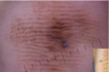
    Figure 1:  A fibrillar pattern with irregular black dots in the center (arrows) and
a parallel ridge pattern in the periphery were detected in dermoscopy.
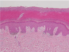
    Figure 2:  Proliferation of atypical melanocytes is distinct in the crista profunda
intermedia. A small amount of melanin pigment can be seen in the cornified
layer arranged in a slanting.
References
- Saida T, Oguchi S, Miyazaki A. Dermoscopy for acral pigmented skin lesions. Clin Dermatol. 2002; 20: 279-285.
- Minagawa A, Omodaka T, Okuyama R. Melanomas and Mechanical Stress Points on the Plantar Surface of the Foot. N Engl J Med. 2016; 374: 2404- 2406.
- Kiyohara T, Satoh S, Yasuta M, Kumakiri M. Irregular fibrillar pattern is an artifactual expression of parallel ridge pattern on the pressure-loaded area of the sole: the efficacy of oblique view dermoscopy. J Dermatol. 2012; 39: 927-928.