
Special Article - Diagnosis and Usefulness of Dermoscopy
Austin J Dermatolog. 2017; 4(2): 1077.
Atypical Pseudonetworks in Pigmented Facial Macules: A Report of Two Cases
Kurita R, Yamamoto Y, Matsue H and Togawa Y*
Department of Dermatology, Chiba University of Graduate School of Medicine, Japan
*Corresponding author: Yaei Togawa, Department of Dermatology, Chiba University of Graduate School of Medicine, 1-8-1, Inohana, Chuo-ku, Chiba, 260-8670, Japan
Received: September 11, 2017; Accepted: October 04, 2017; Published: October 11, 2017
Abstract
We report two rare cases of a black facial macule showing atypical pseudonetwork on dermoscopy. Case 1 was a reticulated type of seborrheic keratosis and case 2 was a superficial Basal Cell Carcinoma (BCC). In general, pseudonetwork is seen in benign or malignant melanocytic lesions. Specifically, an atypical pseudonetwork is known as a hallmark of lentigo maligna and lentigo maligna melanoma. An atypical pseudonetwork is composed of four elements: asymmetrical pigmented follicular openings, rhomboidal structures, annular granular structures, and a gray pseudonetwork. We found no case report of a reticulated-type seborrheic keratosis showing asymmetric pigmented follicular openings with a dark brown rim, as was seen in our case. Furthermore, reticular lines including a pigment network and pseudonetwork are important features, excluding pigmented BCC, and only one case of superficial BCC with an atypical pseudonetwork has been reported. In Case 2, pseudonetwork made of blue-gray network-like pigmentation on pink whitish structureless areas could be seen. It is important to know the diagnostic pitfalls of non-melanocytic pseudonetwork on dermoscopy, although it has useful features for diagnosis of facial pigmented lesions.
Keywords: Atypical pseudonetwork; Basal cell carcinoma; Dermoscopy; Lentigo maligna; Lentigo maligna melanoma; Seborrheic keratosis
Case Presentation
Lentigo Maligna (LM) or Lentigo Maligna Melanoma (LMM) is a type of melanoma that appears predominantly on the face and sun-exposed areas, as large pigmented macules. The presence of a facial atypical pseudonetwork on dermoscopy has been reported to be one of the most important diagnostic clues for LM or LMM [1]. However, with pseudonetwork it is sometimes difficult to distinguish between benign and not benign, as well as between melanocytic or not melanocytic features. Here, we report two cases with a pigmented facial macule with an atypical pseudonetwork that can help to differentiate it from melanoma.
Case 1
An 81-year-old female patient presented with a lesion on her right cheek that appeared 10 years ago. Upon clinical examination, it appeared as a brown macule with multiple shades of maroon, measuring 10 x 8mm (Figure 1A). Dermoscopic examination revealed a brown structureless area free of hair follicles that formed a pseudonetwork with a sharp border (Figure 1B). The pseudonetwork seemed to be atypical, because some asymmetric pigmented follicular openings also could be seen in the lesion as a hyperpigmented rim. Moreover, in the 4 o’clock area of the border, there was an eccentric darker brown pseudonetwork. We hypothesized that the lesion was a solar lentigo because of its structureless area and sharp border. However, we could not rule out LMM completely. We performed a histologic examination of the center of the lesion and of the eccentric pigmented border of the lesion. In the center of the lesion, tumor nests with melanin infiltrated into the upper dermis from the basal layer of the flat epidermis. The tumor nests had two layers of basaloid cells containing melanin that avoided the hair follicles, although they showed uneven melanosis around the follicles (Figure 2A). In the eccentric darker area on the border, tumor nests infiltrated deeper into the dermis and coalesced with each other (Figure 2B). The tumor nests were made of basaloid cells arranged in articulated pattern, and some pseudohorn cysts were also seen. Finally, we diagnosed the lesion as an early stage of reticulated-type seborrheic keratosis.
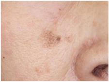
    Figure 1A:  A brown macule with variable shades of brown, measuring 10 x
8mm.
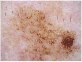
    Figure 1B:  A brown structureless area, free of hair follicles that formed
an atypical pseudonetwork with a sharp border. Asymmetric pigmented
follicular openings as a hyperpigmented dark brown rim could be seen in the
center. In the 4 o’clock region of the border, there was an eccentric darker
pseudonetwork.
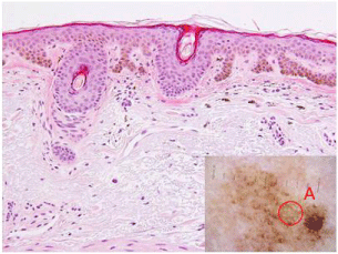
    Figure 2A:  Tumor nests had two layers of basaloid cells containing melanin
when seen in the tissue section of circle “A”. They avoided hair follicles,
nevertheless showing uneven melanosis around the follicles.
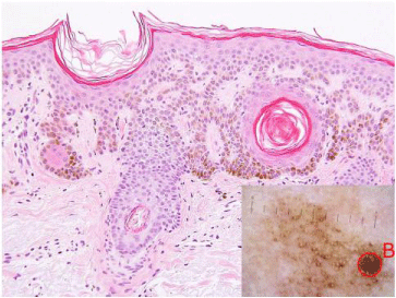
    Figure 2B:  In this area, tumor nests infiltrated deeper into the dermis and
coalesced with each other in the tissue section of circle “B”. Basaloid cells
showed a reticulated pattern, and some pseudohorn cysts were also seen.
Case 2
A 60-year-old male patient presented with a black macule on the left side of his forehead that appeared approximately three years ago. He stated that the lesion had not enlarged in the past several months. Upon clinical examination, the lesion appeared as an irregularshaped black macule, measuring 9 x 5mm (Figure 3A). Dermoscopic examination of the lesion revealed an atypical pseudonetwork with various shades of gray-black and follicles in pseudonetwork were replaced by pink whitish structureless areas (Figure 3B). On the right side of the lesion, asymmetric pigmented follicular openings were seen around hair follicles. On the left side, rhomboidal structures or a gray pseudonetwork in addition to annular-granular structures and gray dots could be seen. Some leaf-like areas and spork-wheel areas could be recognized in periphery and a few mlia-like cysts were also found. We could not rule out LM or LMM because of the atypical pseudonetwork on dermoscopy although some of the clues for BCC were detected; thus, we performed an excisional biopsy.
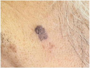
    Figure 3A:  An irregular-shaped black macule with different colors, measuring
9 x 5mm.
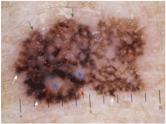
    Figure 3B:  A black pseudonetwork with a partially blue-gray color avoided
the hair follicles. Pink whitish structureless areas replaced hair follicles (blue
arrows). On the right side of the lesion, asymmetric pigmented follicular
openings were seen around hair follicles (yellow dotted circle). On the left
side, rhomboidal structures or a gray pseudonetwork in addition to annulargranular
structures and gray dots could be seen (purple dotted circle). Some
leaf-like areas (green arrows) and spork-wheel areas (white arrows) could be
recognized in periphery and a few mlia-like cysts were also found (red arrow).
The histologic examination showed that the tumor infiltrated into the upper dermis from the basal layer under low magnification (Figure 3C). The infiltrated tumor nests coalesced with each other in the upper dermis, avoiding hair follicles, as seen under high magnification (Figure 3D). The tumor cells containing melanin were basaloid and arranged in a palisading pattern. Inflammatory cells infiltrated moderately with melanophages, and mild fibrosis was also seen. We diagnosed this patient with superficial Basal Cell Carcinoma (BCC).
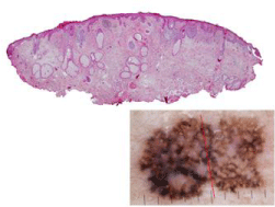
    Figure 3C:  The tumor infiltrated from the flattened epidermis into the upper
dermis. Hair follicles were replaced by tumor nests or flattened epidermis.
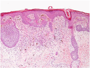
    Figure 3D:  The infiltrated tumor nests coalesced with each other in the
upper dermis, avoiding hair follicles. The basaloid tumor cells containing
melanin were arranged in a palisading pattern. Inflammatory cells infiltrated
moderately with melanophages, and mild fibrosis was seen.
Discussion
A pseudonetwork was defined as a blue-gray to brown diffuse pigmentation formed by the confluence of annular–granular structures surrounding the follicular openings [2]. The typical pseudonetwork that results from the anatomical architecture of the facial skin owing to the flattened rete ridges with pigmentation that avoids the hair follicles is a diagnostic clue for melanocytic lesions and solar lentigo in the facial area, and can exclude BCC in general. Furthermore, atypical pseudonetworks, which are composed of four elements, asymmetric pigmented follicular openings, rhomboidal structures, annular granular structures, and a gray pseudonetwork, are well-known as the hallmark of LM/LMM [3]. In our 2 cases, asymmetric pigmented follicular openings stood out in case 1, and all four findings, especially rhomboidal structures and annular granular structures, were seen in case 2. A pseudonetwork pattern characterized by distribution of the pigment in the proximity of the hair follicles was observed in two of 14 (14.3%) BCCs [4]. The pseudonetworks of the reported case were composed of a blue-gray to brown homogeneous pigmentation, multiple blue-gray globules, and leaf-like areas. In our case, the pseudonetwork was made of blue-gray network-like pigmentation, and follicles in the pseudonetwork were replaced by pink whitish structureless areas. These features represent the components of superficial tumor nests adjacent to epidermis, and histopathologically, we could find a similar case of superficial type BCC with an atypical pseudonetwork [5]; however, we thought this dermoscopic feature of BCC was rather rare. We supposed that other dermoscopic clues for BCC might be helpful in ruling out melanoma in such case. In addition, several lesions such as Lichenplanus- Like Keratosis (LPLK: a kind of irritated seborrheic keratosis) and Pigmented Actinic Keratosis (PAK), can mimic facial LM/LMM [6-8]. However, the common LM/LMM findings of asymmetrical follicular openings and a hyperpigmented rim of follicular openings are very rarely reported in LPLK and PAK [9]. We conjectured the reticulated type of seborrheic keratosis may show sometimes an asymmetrical follicular opening with dark brown rim on dermoscopy, as seen in our case. In a recent multivariate analysis, the most potent predictors of LM were gray rhomboids (six fold increased probability of LM), non evident follicles (four fold) and intense pigmentation (two fold) [10]. Our case lacked gray rhomboids or intense pigmentation around follicules and non evident follicles. Therefore, the perifollicular color and follicular obliteration seems to be the important clue for differentiating LM with other pigmented lesions.
Conclusion
We report two rare cases of a black facial macule showing atypical pseudonetwork on dermoscopy. It is important to know the diagnostic pitfalls of non-melanocytic pseudonetwork on dermoscopy, although it is contains useful features for the diagnosis of facial pigmented lesions. An early stage of reticulated-type seborrheic keratosis has the potential of showing asymmetric pigmented follicular openings made of dark brown rim. In addition, superficial BCC, although rare, may show a pseudonetwork made of blue-gray network-like pigmentation on pink whitish structureless areas.
References
- Peris K, Micantonio T, Piccolo D, Concetta M. Dermoscopic features of actinic keratosis. Jddg. 2007; 5: 970-975.
- Schiffner R, Schiffner-Rohe J, Vogt T, Landthaler M, Wlotzke U, Cognetta AB, et al. Improvement of early recognition of lentigo maligna using dermatoscopy. J Am Acad Dermatol. 2000; 42: 25-32.
- Argenziano G, Soyer HP, Chimenti S, Talamini R, Corona R, Sera F, et al. Dermoscopy of pigmented skin lesions: results of a consensus meeting via the Internet. J Am Acad Dermatol. [Consensus Development Conference Research Support, Non-U.S. Gov’t Review]. 2003; 48: 679-693.
- Gulia A, Altamura D, De Trane S, Micantonio T, Fargnoli MC, Peris K. Pigmented reticular structures in basal cell carcinoma and collision tumours. Br J Dermatol. 2010; 162: 442-444.
- Benez MDV, Silva ALFd, Veradino GC, Silva SCMC. Pigmented basal cell carcinoma mimicking a malignant lentigo melanoma in black female patient. Surg Cosmet Dermatol. 2011; 3: 173-175.
- Akay BN, Kocyigit P, Heper AO, Erdem C. Dermatoscopy of flat pigmented facial lesions: diagnostic challenge between pigmented actinic keratosis and lentigo maligna. Br J Dermatol. 2010; 163: 1212-1217.
- Bugatti L, Filosa G. Dermoscopy of lichen planus-like keratosis: a model of inflammatory regression. J Eur Acad Dermatol Venereol. 2007; 21: 1392- 1397.
- Zaballos P, Ara M, Puig S, Malvehy J. Clinical and dermoscopic image of an intermediate stage of regressing seborrheic keratosis in a lichenoid keratosis. Dermatol Surg. 2005; 31: 102-103.
- Kittler H, Marghoob AA, Argenziano G, Carrera C, Curiel-Lewandrowski C, Hofmann-Wellenhof R, et al. Standardization of terminology in dermoscopy/ dermatoscopy: Results of the third consensus conference of the International Society of Dermoscopy. J Am Acad Dermatol. 2016; 74: 1093-1096.
- Lallas A, Tschandl P, Kyrgidis A, Stolz W, Rabinovitz H, Cameron A, et al. Dermoscopic clues to differentiate facial lentigo maligna from pigmented actinic keratosis. Br J Dermatol. 2016; 174: 1079-1085.