Special Article - Diagnosis and Usefulness of Dermoscopy
Austin J Dermatolog. 2017; 4(3): 1079.
Dermoscopic Features in Fibroepithelioma of Pikus on the Hand: A Case Report
Aoyagi N, Nakano M, Matsue H and Togawa Y*
Department of Dermatology, Chiba University of Graduate School of Medicine, Japan
*Corresponding author: Yaei Togawa, Department of Dermatology, Chiba University of Graduate School of Medicine, 1-8-1, Inohana, Chuo-ku, Chiba, 260-8670, Japan
Received: September 11, 2017; Accepted: November 28, 2017; Published: December 05, 2017
Abstract
Fibroepithelioma of Pinkus (FeP) is generally regarded as a rare variant of Basal Cell Carcinoma (BCC) with distinct clinical and histopathological features, although some researchers have discussed for its classification as a trichoblastoma. Clinically FeP presents as a solitary, soft, skin colored or slightly gray-brown, and polypoid or papillomatous tumor. It is mainly located on the trunk, although it is sometimes seen in the lower extremities and the head and neck. The dermoscopic features of FeP that have been reported include fine arborizing vessels, ulceration, and white streaks in some cases. In addition, graybrown structureless areas and variable numbers of gray-blue dots are found in the pigmented cases. The cases of FeP occurring on the hand are quite rare, and we report the first such case, describing the details of dermoscopic findings. An 83-year-old woman presented with a 3 year history of a solitary, domeshaped, pinkish nodule on the lateral side of the left hand. It developed after a skin burn 3years ago, and enlarged progressively. Dermoscopic examination revealed arborizing vessels, white structureless areas with crystal lines, and an ulceration in the center of the nodule. Histopathologically, the tumor nests in the upper dermis consisted of basaloid cells arranged in anastomotic cords with surrounding fibrous stroma. We diagnosed as the patient as having FeP. There has been no recurrence for 2 years after surgery.
Keywords: Fibroepithelioma of pinkus; Hand; Arborizing vessels; White structureless area
Case Presentation
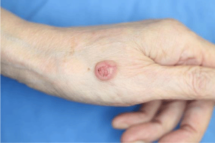
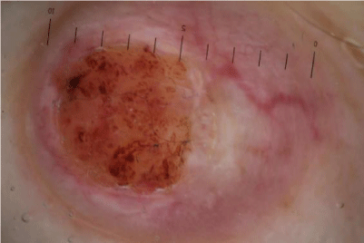
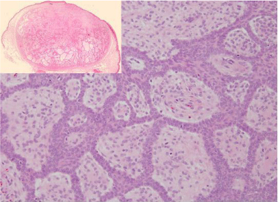
An 83-year-old woman who had no past or family history of irrelevant diseases presented with 3 years history of a solitary, domeshaped, pinkish nodule on the lateral side of the left hand. The nodule occurred after a skin burn 3 years ago and had enlarged gradually. Physical examination revealed a 20 × 10 × 2-mm sized domeshaped pinkish nodule with regular borders on the hand (Figure 1). Dermoscopic examination showed white structureless areas with crystal lines, arborizing vessels, and ulceration with yellow crust in the center of the nodule (Figure 2). Total excision was performed with a 2-mm margin. Histopathologically, the tumor was located in the mid to deep dermis partly connected to the epidermis. The tumor nests consisted of basaloid cells arranged in anastomotic cords and columns. The stroma surrounding them was composed of thick collagen fibers and contained many fibroblasts (Figure 3). Based on these findings, we diagnosed it as FeP. There has been no recurrence for 2 years after excision.

    Figure 1:  A 20 × 10 × 2-mm sized dome-shaped pinkish nodule with regular
borders on the lateral side of the hand.

    Figure 2:  Dermoscopic examination showed white structureless areas with
crystal lines, arborizing vessels, and ulceration with yellow crusts in the
center of the nodule.

    Figure 3:  Histopathology, the tumor nests infiltrated from the epidermis to the
deep dermis. They consisted of basaloid cells arranged in anastomotic cords
and columns. The stroma surrounding them was composed of thick collagen
fibers and contained many fibroblasts.
Discussion
Fibroepithelioma of Pinkus (FeP) was first described in 1953[1]. It is considered to be a rare variant of Basal Cell Carcinoma (BCC) with distinct clinical and histopathological features. However, some researchers have discussed its classification as a trichoblastoma [2].
It is commonly known to occur in adult male individuals aged 40-60 years [3]. FeP usually presents in adults older than 50 years, and female individuals may be affected slightly more often than male individuals because 54% of 114 patients were female [4]. Clinically FeP presents as a soft, usually solitary, polypoid or papillomatous well circumscribed tumor showing skin color or a slightly brown-gray color. FeP is mainly located on the trunk (77.55%), followed by the lower extremities (12.20%) and the head and neck (10.20%) [5]. The most affected part was lower back [6].Therefore we found few reports of FeP occurring on the hand.
Dermoscopic features of FeP were first reported in 2005 by Zalaudek et al. In their largest case series, the lesions were usually red to light brown in color, with fine arborizing vessels (either alone or associated with dotted vessels). In nine of 10 cases, white streaks could be seen. In the pigmented cases, gray-brown structureless areas and variable numbers of gray- blue dots were detected [7].
Longo et al. distinguished a hyperpigmented type of FeP from hypopigmented type [8]. The former showed white intersecting lines with a vascular pattern, and the latter showed ulceration, patchy hyperpigmented areas, and brown dots.
Although some dermoscopic findings of FeP have been reported so far, there are no reports of dermoscopic findings of FeP occurring on the hand.
In our case, arborizing vessels and central ulceration with yellow crust and a pinkish background were observed. White structureless areas with crystal lines, which seemed to have resulted from fibrosis of the upper dermis with flattened epidermis, were also seen. We believe that the reason why our case did not show the white intersecting lines was that the nodule occurred on the border of a non-hairy part of the hand. To our knowledge, this is the first report for the dermoscopic findings of FeP occurring on the hand.
Conclusion
This is the first detailed report of the dermoscopic findings of FeP developed on the hand. We believe that arborizing vessels, central ulceration and white structureless areas might be characteristic dermoscopic features of FeP on the lateral side of the hand, which was at the border of the non-hairy part.
References
- Pinkus H. Premalignant fibroepithelial tumors of skin. AMA Arch Derm Syphilol. 1953; 67: 598-615.
- Ellen S. Haddock. Fibroepithelioma of Pinkus Revisited. Dermatol Ther. 2016; 6: 347-362.
- Pan Z, Huynh N, Sarma DP. Fibroepithelioma of Pinkus in a 9-year-old boy: A case report. Cases J. 2008; 1: 21.
- Bowen AR, LeBoit PE. Fibroepithelioma of Pinkus is a fenestrated trichoblastoma. Am J Dermatopathol. 2005; 27: 149-154.
- Longo C, Pellacani G, Tomasi A, Mandel VD, Ponti G. Fibroepithelioma of Pinkus: Solitary tumor or sign of a complex gastrointestinal syndrome. Mol Clin Oncol. 2016; 4: 797-800.
- Katona TM, Ravis SM, Perkins SM, Moores WB, Billings SD. Expression of androgen receptor by fibroepithelioma of Pinkus: evidence supporting classification as a basal cell carcinoma variant. Am J Dermatopathol. 2007; 29: 7-12.
- Morena S, Pujol RM, Sequra S, Gomez-Martin I. Pigmented fibroepithelioma of Pinkus: A potential dermoscopic simulator of malignant melanoma. J Dermatol. 2017; 44: 542-551.
- Longo C, Soyer HP, Pepe P, Casari A, Wurm EM, Guitera P, et al. In vivo confocal microscopic pattern of fibroepithelioma of Pinkus. Arch Dermatol. 2012; 148: 556.