
Short Communication
Austin J Dermatolog. 2021; 8(1): 1094.
Whole Body FDG PET-CT in the Evaluation and Management of Cardiac Sarcoidosis and Isolated Cardiac Sarcoidosis
Djekidel M*
Department of Diagnostic Imaging, Division of Nuclear Medicine and Molecular Imaging, Qatar
*Corresponding author: Mehdi Djekidel, Department of Diagnostic Imaging, Division of Nuclear Medicine and Molecular Imaging, Sidra Medicine, Al-Luqta Street, POBox Number 26999, Doha, Qatar
Received: December 18, 2020; Accepted: January 25, 2021; Published: February 01, 2021
Introduction
Sarcoidosis is a multisystem inflammatory disease defined histologically by the formation of non-caseating granulomas. Cardiac involvement can be seen in up to 5% of cases [1]. These patients are at a high risk for major cardiac events [1]. Diagnosing and monitoring Cardiac Sarcoidosis (CS) is not trivial but can be currently accomplished by a combination of cardiac MRI and cardiac FDG PET-CT scanning [2]. With appropriate patient preparation, cardiac FDG PET has a high sensitivity for detecting cardiac lesions but also extra-cardiac lesions and for monitoring the efficacy of treatment.
Discussion
Current standard clinical practice consists of acquiring dedicated, limited field of view cardiac FDG PET scans. This would cover the heart and immediately adjacent thoracic structures. Although this is of great value and used with great success to diagnose and monitor cardiac sarcoidosis, it fails to assess the patient’s disease in a holistic manner. Still with this limited field of view, several reports including ours have described a high rate of extra-cardiac findings. We reviewed a series of sixty-five PET-CT scans of 54 patients referred for CS evaluation. These were performed between September 2010 and April 2013 at Yale New Haven Hospital and we found extra-cardiac findings were present in 92.3% of scans (n=60). Overall disease distribution is summarized in (Table 1). Highlight some of these extra-cardiac findings (Figures 1-4). 52.3% of these patients were asymptomatic at the time of the scan. CS was diagnosed in 50.8% of patients (n=33). All the patients (100%) with CS had extra-cardiac findings. Additionally, extra-cardiac findings were present in 48.3% of the scans when there was no cardiac involvement. In our cohort, we had no cases of Isolated Cardiac Sarcoidosis (ICS).
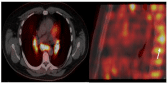
    Figure 1: 52 year old asymptomatic gentleman. FDG PET was negative for
CS, however FDG avid mediastinal and hilar lymphadenopathy was seen in
addition to focal skeletal uptake (white arrow).
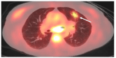
    Figure 2: 55 year old symptomatic lady with palpitations. FDG PET negative
for CS. FDG-avid subcarinal lymph node and left upper lobe nodule seen
(white arrow).
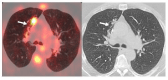
    Figure 3: 69 year old lady presenting with ventricular tachycardia and near
syncope. CS was suspected. However, there was no evidence of CS on
FDG PET. An FDG-avid lung nodule seen incidentally found and it was only
partially visualized (white arrows). The patient underwent a diagnostic chest
CT, which demonstrated a nodule with suspicious features for primary lung
cancer (red arrow). A wedge resection was performed and the pathologic
evaluation revealed organizing pneumonia.
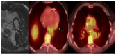
    Figure 4: 73 year old asymptomatic gentleman with known sarcoidosis.
Cardiac MRI study performed 1 year ago showed infero-lateral wall
enhancement (white arrow). FDG PET showed increased uptake in the
same region (green arrow). In addition, he had diffuse FDG uptake along the
thoracic aorta.
![]()
    Percentage


    Mediastinal/Hilar lymphadenopathy
    66.10%


    Proximal stomach/esophagus
    43.10%


    Bone marrow uptake
    35.40%


    Thoracic aorta
    16.90%


    FDG avid lung nodules
    16.90%


    Focal skeletal uptake
    10.80%


    Non-FDG avid lung nodules
    3.10%
Table 1: Distribution of extra-cardiac findings on cardiac sarcoid FDG PET scans.
Baughman et al., reported on findings from the A Case Control Etiologic Study of Sarcoidosis (ACCESS) [3]. This multicenter study evaluated distribution of disease in 736 incident cases with sarcoidosis. One should note that this preceded the widespread use of FDG PET in clinical practice and the true distribution of disease is likely different when highly sensitive techniques such as FDG PET are used. Although ICS is described in the literature with an expected high prevalence ranging from 29-52% of cases of CS [4]. It seems that in the absence of proper staging during the CS workup one may overestimate the cases of ICS. Giudicatti et al., looked into the impact of whole body FDG PET in assessing the true prevalence of ICS [4]. They found it to be much lower at about 9.4%. Juneau et al in their study on the other hand found the prevalence to be even lower at 3.2% [5]. This was much closer to our cohort where we actually found no cases of ICS.
Ishiyama et al., also noted in their study of 16 positive CS cases a high propensity for extra-cardiac disease (81.3%) [1]. Their study also hinted at the potential prognostic value of extra-cardiac disease. Furthermore, extra-cardiac uptake was reported to increase the likelihood of finding CS by Tuominen et al., [6]. Considering that in our cohort all CS patients had extra-cardiac findings it is possible to miss extra-cardiac disease due to a limited field of view or incomplete staging or improper interpretation of the scans. In order not to miss any disease that would increase the diagnostic certainty of having CS as suggested by Tuominen et al., [6], one should strongly consider doing a whole body PET in cases of suspected CS. In these cases, the patient is already being injected with FDG to perform the cardiac study and the additional whole body acquisition mostly adds some additional time to the patient’s acquisition. With newer scanners, this should not be a limiting factor. Higushi et al., [7] reported on a case of large vessel aortitis in a patient with CS as we have also encountered and demonstrated in (Figure 4). Extra-cardiac findings such as this one may have a significant prognostic implication.
Conclusion
Considering the high incidence of extra-cardiac findings in patients undergoing a cardiac sarcoid FDG PET scan, it may seem advisable to perform a whole body FDG PET acquisition in order to properly assess these patients. This would have a high impact for accurate initial staging, monitoring of treatment, assessing differential treatment responses in different aspects of the disease. This also allows for more adequate biopsy sites selection and a more holistic evaluation of the patient.
References
- Ishiyama M, Soine LA, Vesselle HJ. Semi-quantitative metabolic values on FDG PET/CT including extracardiac sites of disease as a predictor of treatment course in patients with cardiac sarcoidosis. EJNMMI Res, 2017; 7: 67.
- Soejima K, Yada H. The work-up and management of patients with apparent or subclinical cardiac sarcoidosis: with emphasis on the associated heart rhythm abnormalities. J Cardiovasc Electrophysiol, 2009; 20: 578-583.
- Baughman RP, Teirstein AS, Judson MA, Rossman MD, Yeager H, Bresnitz EA, et al. Clinical characteristics of patients in a case control study of sarcoidosis. Am J Respir Crit Care Med, 2001; 164: 1885-1889.
- Giudicatti L, Marangou J, Nolan D, Dembo L, Baumwol J, Dwivedi G. The Utility of Whole Body (18) F-FDG PET-CT in Diagnosing Isolated Cardiac Sarcoidosis: The Western Australian Cardiac Sarcoid Study. Heart Lung Circ. 2020; 29: e1-e6.
- Juneau D, Nery P, Russo J, de Kemp RA, Leung E, Beanlands RSB, et al. How common is isolated cardiac sarcoidosis? Extra-cardiac and cardiac findings on clinical examination and whole-body (18) F-fluorodeoxyglucose positron emission tomography. Int J Cardiol. 2018; 253: 189-193.
- Tuominen H, Haarala A, Tikkakoski A, Kahonen M, Nikus K, Sipila K. 18-FDGPET in a patient cohort suspected for cardiac sarcoidosis: Right ventricular uptake is associated with pathological uptake in mediastinal lymph nodes. J Nucl Cardiol. 2020; 27: 109-117.
- Higuchi Y, Kimoto Y, Tanoue R, Tokunou T, Tomonari K, Maeda T, et al. Cardiac Sarcoidosis Concomitant with Large-vessel Aortitis Detected by 18F-fluorodeoxyglucose Positron Emission Tomography. Internal Medicine. 2018; 57: 1601-1604.