Short Communication
Austin J Dermatolog. 2021; 8(1): 1095.
FDG PET Scanning in Hemophagocytic Lymphohistiocytosis
Djekidel M*
Department of Diagnostic Imaging, Division of Nuclear Medicine and Molecular Imaging, Qatar
*Corresponding author: Mehdi Djekidel, Department of Diagnostic Imaging, Division of Nuclear Medicine and Molecular Imaging, Sidra Medicine, Al-Luqta Street, POBox Number 26999, Doha, Qatar
Received: December 18, 2020; Accepted: January 25, 2021; Published: February 01, 2021
Introduction
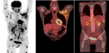
Hemophagocytic Lymphohistiocytosis (HLH) is a rare disorder. It progresses rapidly and can be life-threatening. Prompt initiation of treatment is critical for survival. Untreated, patients survive only a few months. These patients have multisystem involvement and may develop multi-organ failure. The goal of therapy for patients with HLH is to suppress life-threatening inflammation. After induction, patients who are recovering are weaned off therapy, while those who are not improving are continued on therapy as a potential bridge to stem cell transplantation. Monitoring treatment response is critical as treatment escalation may be warranted if the patient is not improving. The distinction between chemotherapy toxicity and worsening disease may be difficult to make clinically. Quite a variety of clinical, biochemical and immunological biomarkers are used at diagnosis and to assess treatment response. The response to initial therapy is a major factor in determining the need for additional therapy including Hematopoietic Cell Transplant (HCT). Response to induction therapy is monitored by assessing the patient clinically and using HLH disease-specific markers. Although this approach has some success, it is sometimes confounded by a new infection or treatment toxicity. Imaging can provide an additional layer of accuracy in the initial evaluation of HLH patients as well as their monitoring and follow-up. 18F-FDG PET scans are highly sensitive and can be quite helpful in the management of these patients (Figure 1).

    Figure 1: 20 yo gentleman with HLH. FDG PET scan showing multiple FDG
avid lesions noted involving muscles and subcutaneous tissues in the face,
arms, chest, abdomen and lower extremities. FDG avid lung nodules and mild
diffuse bone marrow uptake is also noted. Ferritin levels elevated at 13,500.
Patient passed away within a few weeks of initial diagnosis.
Discussion
FDG PET-CT is an established management tool in a variety of cancers, however in noncancerous entities clinicians are more hesitant to use. That said FDG PET has been described as having a high impact in the care of HLH patients. Several authors have described the value of FDG PET scans in delineating the extent of disease in HLH as well as determining the severity/aggressiveness of disease involvement [1-6]. Wang et al, described the added value of FDG PET scans in delineating lymphoma lesions and managing lymphoma associated HLH [7]. On the other hand Yang et al, raised the concern of using FDG PET as a tool to assess bone marrow involvement by lymphoma in lymphoma associated HLH as HLH related bone marrow changes may frequently be a confounding factor [8]. Diffuse bone marrow uptake in HLH has also been described by several other authors [1, 8-11].
Additionally, HLH associated cancers can be detected by FDG PET scans and guide biopsies [4]. In 45 HLH patients Jigang described the distribution and pattern of uptake in secondary HLH cases including rheumatological systemic diseases, infections, and lymphoma [5,12]. Considering HLH is even less common in children and early diagnosis is critical in management and improving morbidity and mortality one should consider using FDG PET in cases of fever of unknown origin or other nonspecific presentations including in pediatrics. Adequate, early assessment of these patients is critical [2,13,14]. FDG PET can detect disease early and is a better predictor of prognosis and outcomes than clinical or laboratory biomarkers [1,6]. Therefore, one would also postulate that it would be a beneficial tool in treatment monitoring.
Conclusion
HLH is a serious life-threatening disease. FDG PET can play a major role in early disease assessment, prognosis, biopsy guidance, the evaluation of secondary causes and treatment monitoring. Larger multicenter studies are required for better guidance in this rare and fatal disease.
References
- Ersahin D, Djekidel M. Multimodality imaging with F18 FDG PET in a patient with Hemophagocytic Lymphohistiocytosis (HLH). J Nucl Med. 2011; 52: 1010.
- Li F, Su M, Tian R, Miller J. Hemophagocytic lymphohistiocytosis-18F-FDG PET/CT findings in children. J Nucl Med. 2015; 56: 1711.
- Recht H, Solnes L, Merrill S, Siegelman S. 18-FDG PET/CT findings in hemophagocytic lymphohistiocytosis: a pictorial review. J Nucl Med. 2020; 61: 1140.
- Kim J, Song HC, Kang SR, Yoo SW, Oh JR, Chong A, et al. Clinical usefulness of F-18 FDG PET/CT in patients with hemophagocytic lymphohistiocytosis. J Nucl Med. 2011; 52: 1875.
- Yamashita H, Kubota K, Takahashi Y, Minamimoto R, Morooka M, Kaneko H, et al., Clinical value of (1) (8) F-fluoro-dexoxyglucose positron emission tomography/computed tomography in patients with adult-onset Still’s disease: a seven-case series and review of the literature. Mod Rheumatol. 2014; 24: 645-650.
- Kim J, Yoo SW, Kang SR, Bom HS, Song HC, Min JJ. Clinical implication of F-18 FDG PET/CT in patients with secondary hemophagocytic lymphohistiocytosis. Ann Hematol, 2014; 93: 661-667.
- Wang J, Wang D, Zhang Q, Duan L, Tian T, Zhang X, et al. The significance of pre-therapeutic F-18-FDG PET-CT in lymphoma-associated hemophagocytic lymphohistiocytosis when pathological evidence is unavailable. J Cancer Res Clin Oncol. 2016; 142: 859-871.
- Yang YQ, Ding CY, Xu J, Fan L, Wang L, Tian T, et al. Exploring the role of bone marrow increased FDG uptake on PET/CT in patients with lymphomaassociated hemophagocytic lymphohistiocytosis: a reflection of bone marrow involvement or cytokine storm? Leuk Lymphoma. 2016; 57: 291-298.
- Yiu CR, Kao YH, Phipps C, Tan D. Positron emission tomography findings in patients with lymphoma-associated haemophagocytic syndrome. Singapore Med J. 2011; 52: e156-e159.
- Harada S, Shinohara T, Naruse K, Machida H. Diffuse 18F-fluorodeoxyglucose accumulation in the bone marrow of a patient with haemophagocytic lymphohistiocytosis due to Hodgkin lymphoma. BMJ Case Rep. 2016; 2016: bcr2016217555.
- Corapcioglu F, Oncel S, Berberoglu K, Klc SC, Aydogan A, Dogan S. False positivity of FDG-PET during hemophagocytic lymphohistiocytosis in a child with Hodgkin lymphoma in remission. J Pediatr Hematol Oncol. 2009; 31: 74-75.
- Yang J. The 18F-FDG PET/CT is helpful in determining the cause and extent of secondary hemophagocytic lymphohistiocytosis. J Nucl Med. 2015; 56: 1424.
- Koc ZP, Akarsu S, Balci T, Unal K. PET/CT images of a patient with haemophagocytic lymphohistiocytosis. BMJ Case Rep. 2012; 2012: bcr0320126026.
- Jasper N, Dabritz J, Frosch M, Loeffler M, Weckesser M, Foell D. Diagnostic value of [(18) F]-FDG PET/CT in children with fever of unknown origin or unexplained signs of inflammation. Eur J Nucl Med Mol Imaging. 2010; 37: 136-145.