Case Report
Austin J Dermatolog. 2022; 9(1): 1102.
Congenital Infection with Cytomegalovirus Presented as Blueberry Muffin Syndrome in a Preterm Newborn: A Case Report
Farhadi R*
Associate Professor of Neonatology, Department of Pediatrics, Mazandaran University of Medical Sciences, Sari, Iran
*Corresponding author: Roya Farhadi, Associate Professor of Neonatology, Division of neonatology, Department of Pediatrics, Boo Ali Sina Hospital, Pasdaran Boulevard, Mazandaran University of Medical Sciences, Sari, Iran
Received: June 09, 2022; Accepted: July 06, 2022; Published: July 13, 2022
Abstract
Blueberry muffin baby, describes a neonate with multiple purpuras, associated with several conditions in which extra blood is created in the skin. In this report, a premature baby is introduced who had skin lesions in the form of blueberry muffin at birth, and the related differential diagnosis and the final etiology of this baby will be reviewed.
Keywords: Congenital; Cytomegalovirus; Infections; Newborn; Purpura; Skin
Introduction
Blueberry muffin rash is consistent with extramedullary hematopoiesisandrefers to multiple purpuras and red-to-violaceous macules, papules, or nodules present within 2 days of life [1-3]. It is a nonspecific and rare clinical presentation in neonates [4,5]. The term “blueberry muffin baby” was primarily used to describe the cutaneous manifestations of congenital rubella in newborns during the American epidemic in the 1960s [6,7]. The lesions tend to occur on the head, neck, and trunk [8]. The differential diagnosis of blueberry muffin baby includes congenital infections from the TORCH (toxoplasmosis, others (hepatitis B or others), rubella, cytomegalovirus, herpes simplex virus) group, dermal extramedullary hematopoiesis or severe hemolysis, many early-onset malignancies or infiltrative neoplastic lesions of the skin, and cutaneous vascular anomalies [9].
In this casereport, a newborn with congenital Cytomegalovirus (CMV) infection manifested as a blueberry muffin baby at birth is described.
Case Presentation
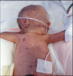
A preterm male newborn was admitted at birth to NICU (neonatal intensive care unit) forrespiratory distress and presenting disseminated violaceous cutaneous lesions. The baby was born preterm weighing 1300 grams at 35 weeks of gestation by cesarean section with the prenatal diagnosis of intrauterine growth retardation. Maternal serological tests for TORCH performed in the first trimester of pregnancy did not show abnormal results and were negative. The lesions were red to violaceous annular macules in size 2- 5 mm (Figure 1). On initial examination, abdominal distention with hepatosplenomegaly (which was also confirmed by ultrasound) and a heart murmur in cardiac auscultation (which was reported as a patent ductus arteriosus by echocardiography) was revealed. The newborn blood laboratory exams showed thrombocytopenia, elevated transaminases, direct hyperbilirubinemia, and coagulopathy.A cerebral ultrasound examination showed periventricular hyper echogenicity. We were not able to perform a brain CT scan due to the unstable condition of the baby. The placental anatomical exam showed features suggestive of congenital infection. It was not possible to get a skin biopsy due to parental dissatisfaction. With early suspicion of congenital viral infections; mother and newborn TORCH screening were requested to find out the causative virus. The results showed that the immunoglobulin M (IgM) titer for CMV (cytomegalovirus) was very positive in the newborn and the level of immunoglobulin G (IgG) antibody was higher than mother.After that, the Polymerase Chain Reaction (PCR) test results of the neonatal saliva and urine were positive for the CMV,and the inclusion body of the virus was observed in the samples. On the sixth day of life, based on clinical, biochemical, and PCR findings, a diagnosis of a blueberry muffin baby with CMV hepatitis was made and intravenous ganciclovir 100 mg per day was initiated for the baby. Within 4 weeks, the skin lesions were faded and ganciclovir continued for 6 weeks. Results of ophthalmologic exams and fundoscopy were within normal limits. Early hearing screening of the infant showed abnormal left ear otoacoustic emission testing, which was scheduled to be re-examined with more specialized tests. The infant was discharged at the age of 50 days with the recommendation of an outpatient visit for follow-up.

Figure 1: Disseminated violaceous lesions at birth.
Discussion
In this report of a blueberry muffin baby, the final diagnosis was congenital CMV infection. The diagnosis of CMV infection was confirmed according to positives erologic results and detection of the virus by PCR technique in urine and saliva. As the blueberry muffin baby has the potential for serious systemic complications, a cause must be considered for all newborns [10]. Various causes have been introduced for blueberry muffin syndrome including intrauterine infections such as rubella, toxoplasmosis, cytomegalovirus, chicken pox, chlamydia, HIV, parvovirus, Epstein Barr virus, and syphilis [11]. Shah et al reported a case of CMV hepatitis presented with a blueberry muffin [3]. Hematologic disorders are another cause of this lesion such as spherocytosis, alloimmunization, twin to twin transfusion syndrome, and fetomaternal transfusion [3]. Daum et al reported a blueberry muffin baby due to hereditary spherocytosis [12]. In some cases, skin lesions may be created because of inborn errors of metabolic disorders. Wagner et al showed that mevalonic aciduria should be considered as a differential diagnosis of blueberry muffin baby [11]. Neoplastic disorders including leukemia, neuroblastoma, and rhabdomyosarcoma are one of the important causes of blueberry muffin baby syndrome. Langerhans cell histiocytosis (LCH) may affect single or multiple organ systems. Skin involvement in LCH has been reported to occur in more than half of the babies at birth [13]. Congenital vascular lesions such as multiple hemangiomas of infancy may be classified as one of the causes of blueberry muffin baby syndrome [3].
Extramedullary hematopoiesis occurs normally during embryologic development in several organs, including the dermis and this activity continues until the fifth month of gestation. Although the exact mechanism of prolonged dermal erythropoiesis in blueberry muffin syndrome is unknown, the presence of lesions at birth shows a postnatal expression of this normal fetal extramedullary hematopoiesis [1].
In this report, the diagnosis was made according to the review of the pregnancy history and prenatal laboratory exams with a special focus on positive serology’s of CMV infection associated with hematological alterations, hepatosplenomegaly, and cholestasis and a definite diagnosis was confirmed by observing CMV shedding in baby’s fluids which detected by polymerase chain reaction. So, CMV infection should be investigated carefully in these babies for early commencement of therapy and prevention of comorbidities.
Conclusion
This case presentation is proposed to highlight that although blueberry muffin baby is a rare manifestation at birth, congenital cytomegalovirus infection should be considered in the differential diagnosis of this syndrome.
Declarations
Consent for publication: Written informed consent was obtained from the patient‘s parents for publication of this case report and any accompanying image.
Competing interest: There is no competing interest.
References
- Shetty G, Kalyanshetti R, Khan HU, Hegde P. Blueberry muffin rash at birth due to congenital rubella syndrome. Indian J Paediatr Dermatol. 2013; 14: 73 5.
- Martins S, Rocha G, Silva G, Calistru A, Pissarra S, Guimarães H. [Blueberry muffin baby. A rare presentation of congenital cytomegalovirus infection]. Acta medica portuguesa. 2011; 24: 703-8. doi:10.20344/AMP.1535.
- Shah VH, Rambhia KD, Mukhi JI, Singh RP. Blueberry Muffin Baby with Cytomegalovirus Hepatitis. Indian Dermatology Online Journal. 2019; 10(3): 327. doi:10.4103/idoj.IDOJ_291_18.
- Chyzynski B, Matysiak M. Blueberry muffin baby syndrome – review paper. Nowa Pediatr. 2019; 23: 34-9.
- Kaleta K, Klosowicz A, Jusko N, Kapiñska-Mrowiecka M. Blueberry muffin baby syndrome. A critical primary sign of systemic disease. Postepy Dermatol Alergol. 2022; 39(2): 418-20.
- Barnett HL, Einhorn AH. Paediatrics. 14th ed. Appleton- Century- Crofts, New York 1968; 742.
- Mehta V, Balachandran C, Lonikar V. Blueberry muffin baby: a pictoral differential diagnosis. Dermatology online journal. 2008; 14(2): 8.
- Bagna R, Bertino E, Rovelli I, Peila C, Giuliani F, Occhi L, et al. Benign transient blueberry muffin baby. Minerva pediatrica. 2010; 62(3): 323-7.
- Isaacs H. Cutaneous Metastases in Neonates: A Review. Pediatric Dermatology. 2011; 28(2): 85-93. doi:10.1111/j.1525-1470.2011.01372.x.
- Karmegaraj B, Vijayakumar S, Ramanathan R, Samikannu R. Extramedullary haematopoiesis resembling a blueberry muffin, in a neonate. BMJ Case Reports. 2015; 2015: bcr2014208473. doi:10.1136/bcr-2014-208473.
- Wagner R, Bellettini SV, Bandeira M, Gubert EM, Schmitz ML. Mevalonic aciduria as a differential diagnosis of blueberry muffin baby. J Neonatal Biol. 2016; 5: 225.
- Daum LM, Sklar LR, Mehregan DR. Blueberry muffin rash secondary to hereditary spherocytosis. Cutis. 2018; 101(2): 111-114.
- Hansel K, Tramontana M, Troiani S, Benedictis DD, Bianchi L, Cucchia R, et al. Congenital Self-Healing Langerhans Cell Histiocytosis: A Rare Presentation of Blueberry Muffin Baby “Spectrum”. Dermatopathology. 2019; 6(2): 37-40. doi:10.1159/000499311.