Case Report
Austin Endocrinol Diabetes Case Rep. 2023; 7(1): 1019.
Aldosterone Producing Adenoma Associated Hypercalcemia and Rhabdomyolysis
Shahnaz Ahmad Mir; Pooran Sharma*; Md Ejaz Ala
Department of Endocrinology, GMC, Srinagar, India
*Corresponding author: Pooran Sharma Department of Endocrinology, GMC, Srinagar, India. Email: puran.k.sharma@gmail.com
Received: April 03, 2023 Accepted: May 01, 2023 Published: May 08, 2023
Abstract
Aldosterone producing adenomas are rare in children’s with less than 20 cases reporting in literature present a 15-year female who presenting with polyuria and proximal muscle weakness. Investigations revealed severe hypokalaemia, raised creatinine phosphokinase suggestive of hypokalaemia induced rhabdomyolysis. Further patient’s investigations showed raised aldosterone and suppressed renin and CT abdomen was suggestive of left adrenal adenoma which was removed laparoscopically. We will review all cases in literature of childhood onset aldosteronomas.
Keywords: Aldosteronoma; Hypokalaemia; Rhabdomyolysis; Polyuria
Introduction
It is well known that marked hypokalaemia manifests as muscle weakness, but rhabdomyolysis is a rare presentation of hypokalaemia. The causes of rhabdomyolysis are various; usage of laxatives and diuretics, anorexia, chronic alcoholism, infectious enterocolitis, renal tubular acidosis and aldosteronism has been reported to be possible causes of hypokalaemia rhabdomyolysis [1,2]. However, aldosterone-producing adrenal tumours are extremely rare in children, considering the cause of hypokalaemia. Here, we report a case of primary hyperaldosteronism due to unilateral aldosterone producing adenoma in a 15-year-old girl who developed rhabdomyolysis following hypokalaemia.
Case Report
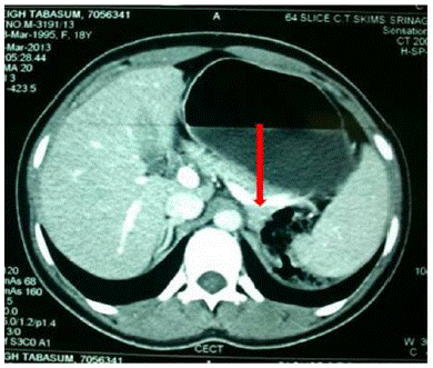
13 year female presented with Polyuria, (documented urine output 7.5 lit], increase thirst for 6 months; severe myalgias and inability to stand from sitting position for 2 months. No history of failure to thrive, or any alternative medicine intake, hyper or hypothyroid symptoms, or any problem with hearing. No significant family history, two other siblings normal. Anthropometry: Height-153cm [10-25%], Weight [48kg]. Puberty completed, no clinical evidence of rickets or any bone disease, Pulse 78 regular B.P-140/90mmHg (both upper limbs), Chest- WNL CVS; WNL, Bulk normal, both upper and lower limbs Tone normal, Power grade 4- in all muscle groups of upper and lower limbs. Gower's sign was positive. Reflexes diminished. Patient serum electrolytes revealed hypokalaemia, metabolic acidosis and hypernatermia. Patient had normal liver and kidney function tests. Serum LDH was 680U/L and CPK of 3320U/L. Patient had calcium of 10.2 and phosphorus of 3.8mg/dl. 2hr urinary calcium was 550mg/day. ECG revealed U waves and USG abdomen showed Anderson cork kidney. Serum vitamin D was 48ng/ml and serum PTH of 110pg/ml. Patients workup for hyperaldosteronism revealed serum aldosterone of 1182pg/ml and renin of 0.013ng/ml/hr. CECT abdomen showed a (1.2×1.8cm) left adrenal mass (Figure 1). Laparoscopic adrenalectomy was done which showed a (2×1.3cm) tumor found in left adrenal gland. Histopathology confirmed left adrenal adenoma (Figure 2). Patient is on follow-up off any treatment for 6 months.

Figure 1:

Figure 2:
Discussion
Hypokalemia is generally associated with neuromuscular symptoms and acid-base disorders. Potassium depletion from any cause may produce muscle weakness, but hypokalaemic rhabdomyolysis is a rare presentation of hypokalaemia. Rhabdomyolysis from any cause lead to increase in free ionised calcium in the sarcoplasm. This increased sarcoplasmic calcium initiates a series of complex intracellular processes leading to the clinical manifestation of rhabdomyolysis [2].
On initial assessment, our patient presented with rhabdomyolysis including weakness of lower limbs, increased creatinine phosphokinase and mild tubulopathy including polyuria and hypokalaemia. The severity of rhabdomyolysis varies from an asymptomatic increase in creatinine phosphokinase to severe complications such as acute renal failure [2] Tubulopathy might be secondary to hypokalaemia or rhabdomyolysis in our patient. Rhabdomyolysis may cause renal tubular damage and tubulopathy either by free radical mediated injury or directly by effect of lipid peroxidation on tubular cells. Chronic hypokalaemia can also induce tubulointerstitial damage consisting of vacuolization of epithelial tubular cells and interstitial fibrosis; called as hypokalaemia nephropathy, which is quite rare [3].
Aldosterone producing adrenocortical tumors are usually benign (adenoma) and adrenocortical carcinomas constitute less than 0.2% of all childhood neoplasms [4]. This differentiation between adenoma and carcinoma is important for prognosis, but is not always easy. MRI has been proposed to have diagnostic value for this differentiation [5].
Primary hyperaldosteronism due to an aldosterone-secreting adenoma associated with hypokalaemia and rhabdomyolysis has been reported in only few adults [6,7]. Samanthi A et al. [6] reported a 42-year-old hypertensive patient who developed acute onset paraparesis and rhabdomyolysis induced by severe hypokalaemia diagnosed an aldosterone-producing adenoma. Hypertension is unusual in children and endocrine causes are very rare, but primary hyperaldosteronism should always be considered in the differential diagnosis [8-10]. Recently, Karaguzel et al [11] Reported a 14 year old girl with primary hyperaldosteronism due to unilateral aldosterone producing adenoma who developed rhabdomyolysis following hypokalaemia. Primary hyperaldosteronism due to an adrenal tumor is rare in childhood; however, it should be considered in the presence of hypokalaemia in a hypertensive child with normal renal function, even if the clinical presentation is unusual, such as severe rhabdomyolysis, of which early recognition is crucial for interventions directed to preserve renal function.
References
- Brumback RA, Feeback DL, Leech RW. Rhabdomyolysis in childhood. A primer on normal muscle function and selected metabolic myopathies characterised by disoederde energy production. Pediatr Clin North Am. 1992; 39: 821-858.
- Chatzizisis YS, Misirli G, Hatzitolios AI, Giannoglou GD. The syndrome of rhabdomyolysis: complications and treatment. Eur J Intern Med. 2008; 19: 568-574.
- Holt SG, Moore KP. Pathogenesis and treatment of renal dysfunction in rhabdomyolysis. Intens Care Med. 2001; 27: 803-811.
- Ciftci AO, Senocak ME, Tanyel FC, Buyukpamukcu N. Adrenocortical tumors in children. J Pediatr Surg. 2001; 36: 549-554.
- Higgins CB, Hricak H. Magnetic Resonance Imaging of Body. New York: Raven Press. 1987; 383.
- Mourad JJ, Milliez P, Blacher J, Safar M, Girerd X. Conn adenoma manifesting as reversible tetraperesis and rhabdomyolysis. Rev Med Interne. 1998; 19: 203-205.
- Chow CP, Symonds CJ, Zodhodne DW. Hyperglycemia, lumbar plexopathy and hypokalemic rhabdomyolysis complicating conn’s syndrome. Can J Neurol Sci. 1997; 24: 67-69.
- Chudler RM, Kay R. Adrenocortical adenoma in children. Urol Clin North Am. 1989; 16: 469-479.
- Rodriquez-Arnao J, Perry L, Dacie JE, Reznek R, Ross RJ. Primary hyperaldosteronism due to an adrenal adenoma in a 14- year old boy. Postgrad Med J. 1995; 71: 104-106.
- Dinleyici EC, Dogruel N, Acikalin MF, Tokar B, Oztelcen B, et al. An additional child case of an aldosterone producing adenoma with an atypical presentation of peripheral paralysis due to hypokalemia. J Endocrinol Invest. 2007; 30: 870-872.
- Karaguzel G, Bahat E, Imamoglu M, Ahmetoglu A, Yildiz K, et al. An unusual case of an aldosterone producing adrenocortical adenoma presenting with rhabdomyolysis. J Peditr Endocr Met. 2009; 22: 1087-1090.