Case Report
Austin Endocrinol Diabetes Case Rep. 2024; 8(1): 1020.
Unilateral Lisch Nodules Singular Clinical Manifestation of Neurofibromatosis 1 Simultaneous Undiagnosed Type 2 Diabetes Mellitus
Nurmamed Serdarov¹; Selbi Hudaýgulyýeva¹; Merjen Myradova¹; Leyli Ovezova¹; Ogulnur Baýrammuhammedova²; Merjen Allaberdyeva³; Mayagozel Zhutdieva¹*
¹International Center of Endocrinology and Surgery, Ashgabat, Turkmenistan
²Arkadag City Health Care Center, Ashgabat, Turkmenistan
³International Center of Neurology, Ashgabat, Turkmeni
*Corresponding author: Mayagozel Zhutdieva International Center of Endocrinology and Surgery, Ashgabat, Turkmenistan. Email: dr.zhutdieva@mail.ru
Received: May 30, 2024 Accepted: June 18, 2024 Published: June 25, 2024
Abstract
Background: Neurofibromatosis type 1 is an autosomal dominant hereditary disease that increases the risk of developing benign and malignant tumors. It allows the growth of tumors along the nerves of bone, skin and brain. Autoimmune disease associated with NF1 can be seen but it is rarely associated neurofibromatosis with diabetes mellitus.
Case Report: We report on a 30-year-old female with undiagnosed type 2 diabetes mellitus and the only manifestation of neurofibromatosis type 1 as unilateral Lisch nodules without other ophthalmological problems. Blood tests revealed hyperglycemia: 18.7 mmol/l (blood glucose concentration: reference range 3.8-6.1 mmol/l), glycated hemoglobin: 10.5% (HbA1c: reference range 4.3-6.0%).
Conclusion: In our case demonstrate of the only clinical manifestation on the iris by neurofibromatosis 1 as Lisch nodules on the right eye identified by routine ophthalmological examination about undiagnosed diabetes mellitus. Both proper diagnosis and treatment of the patient with neurofibromatosis type 1 and undiagnosed diabetes mellitus require cooperation of different specialties. An early multidisciplinary consultation may lead to more effective treatment strategies.
Keywords: Neurofibromatosis type1; Lisch nodules; Diabetes mellitus type 2; Hyperglycemia
Introduction
Neurofibromatosis type 1 is an autosomal dominant disorder, first described by von Recklinghausen in 1882, [1-3], and occurring with an estimated incidence of 1:2000 to 1:3000 individuals of gender, race and ethnicity [4]. Endocrine diseases and neoplasia occurs in patients with NF-1, which include phaeochromocytomas/paragangliomas, primary hyperparathyroidism, gastroenteropancreatic neuroendocrine tumors, thyroid, and other adrenal tumors [2].
The clinical manifestations include cafe au lait spots; cutaneous, subcutaneous, and plexiform neurofibromas; axillary and/or inguinal freckling; Lisch nodules; intracranial gliomas; malignant peripheral nerve sheath tumors; vascular and bone dysplasia [5].
Hamartomas of the iris or melanocytic nevi can be seen and called Lisch nodules. They are variable in size and have a smooth, dome-shaped configuration [6]. The most common ophthalmic manifestation Lish nodules two or more are the one of diagnostic criteria for NF1 [7]. Huson et al. [8] reported that Lisch nodules were present in 95% of their patients and were bilateral in 93%. Numerously, one sided Lisch nodules ensure frequently, as well somatic mosaicism may describe in our case. Iris hamartomas which can be indicative of Neurofibromatosis 1 when multiple, are rarely seen in Neurofibromatosis 2 [9]. One sided numerously Lisch nodules appear frequently same as Neurofibromatosis type 1 as well it can be seen along with diabetes mellitus.
We reported case with rare combination of neurofibromatosis type 1 and type 2 diabetes mellitus.
Case Report
A 30-year old non-obese woman was born with non- consanguineous marriage, she have been at International Center of Endocrinology and Surgery Ashgabat, Turkmenistan reason of she has typical symptoms of diabetes mellitus as polydipsia, polyuria, xerostomia, dry skin, itching of the genital area within one week. She initially visited our clinic and by result of blood examination her blood glucose and glycated hemoglobin (HbA1c) levels found 18.7 mmol/l and 10.5%, respectively. From conversation with an ophthalmologist, we found that mother and her aunt have clinical diagnose of neurofibromatosis type 1 and four consensus criteria for neurofibromatosis exactly neurofibromas, axillary freckling, café-au-lait macules, Lisch nodules.
The patient had not any history of drug allergy, cardiac/renal diseases. She never had similar symptoms in the past. She did not report any family history of diabetes mellitus (parents, two sisters and two brothers were healthy). From her over the past one-year report, we found that she consumed sugar-containing soft drinks.
Have not found any history of visual or auditory disturbances, neurological symptoms, speech impairment and skeletal abnormalities. She was hospitalized in our clinic and have examined by endocrinologist, dermatologist, neurologist, ophthalmologist, otorhinolaryngologist, orthopedist, gynecologist.
Physical Examination
Period of visiting clinic, the general examination of the patient revealed with normal results, and vital signs were stable (pulse rate 70 per minute, respiratory rate 16 per minute, temperature 36.6°C, blood pressure 110/70 mmHg). Anthropometric measurements revealed with weight of 60 kg and a height is 167 cm (BMI: 21.5 kg/m2). Examination of skin revealed with absence of typical clinical signs von Recklinghausen’s disease: no neurofibromas, café-au-lait macules (CALMs), axillary, or inguinal freckling. She had normal vesicular breath sounds in bilateral lung fields and it was negative for any neurological deficits. The skin and mucous membranes were not affected. The abdomen was soft and flat, the liver and spleen were not palpable.
Laboratory Examination
Blood examination revealed high levels of blood sugar: 18.7 mmol/l (RR: 3.8-6.1 mmol/l), glycated hemoglobin: 10.5% (RR: 4.3-6.0%). Blood tests include complete hemogram (total leukocyte, red blood cell and platelet count) except for hemoglobin, coagulogram (APTT, INR, and fibrin degradation product level) and routine blood biochemistry (serum urea, creatinine, the serum levels of triglycerides, low density lipoproteins, high-density lipoproteins and calcium) were within normal limits. Biochemical data are summarized in Table 1.
    Laboratory
    Specimen
    Patient
    Reference range


    Biochemistry


    Proteins total
    serum
    79
    60-78 g/l


    Albumin
    serum
    34.3
    35-50 g/l


    Bilirubin total
    serum
    1.2
    0.1-1.2 mg/dl


    Triglycerides
    serum
    1.0
    0-1.7 mmol/l


    Cholesterol (total)
    serum
    3.8
    3.9- 5.2 mmol/l


    Glucose
    serum
    18.7
    3.8-6.1 mmol/l


    HbA1c
    serum
    10.5
    4.3-6.0%


    Amylase
    serum
    95
    25-125 U/l
Table 1: Laboratory test results.
Opthalmological Examination
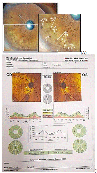
The ophthalmological examination of uncorrected visual acuity was 20/20 on both the eyes. Intraocular pressure determined by non-contact tonometry was 14 mmHg and 16 mmHg in the right and left eye, respectively. Slit-lamp examination revealed with normal anterior eye segments except for the iris. On the anterior surface of her right iris periphery small papules (Figure 1a, b). At the high magnification in the form of grains from light brown to dark brown colour (Figure 1c, d). By direct ophthalmoscopy, there was a clear view on the posterior pole in both eyes with a normally appearing optic disc, retinal vessels, and macula.

Figure 1: Opthalmologiсal clinical dates. (A) Slit-lamp photomicrographs of the anterior segment of the right eye at the first visit. Multiple small, oval, yellow-brown papules. (B) bilaterally optic nerve head topography (OCT; Spectralis, Heidelberg Engineering, Heidelberg, Germany).
Optical coherence tomography (OCT; Spectralis, Heidelberg Engineering, Heidelberg, Germany) demonstrated that the part of the optic disc is not changed for both eyes and are located in green area. The state of the optical fiber layer around the optic disc of both eyes is shown in Figure 1.
Discussion
NF1is a relatively common autosomal dominant disease and the specific gene maps to chromosome 17q11.2. The criteria for diagnosis of NF1, developed by a National Institutes of Health (NIH) Consensus Conference in 1987, which established for routine clinical manifestations of NF1 include cafe-au-lait spots, freckling, generalized cutaneous neurofibroma, Lisch nodules, short stature, optic glioma, and CNS tumors. [10].
Only few reports clarify the association between NF1 and other autoimmune diseases as systemic lupus erythromatosus, mixed connective tissue disease, rheumatoid arthritis, glomerulonephritis, as well as bullous pemphigoid and vitiligo, acromegaly and goiter [11,12].
Lish nodules are the most common ophthalmic manifestation of NF1 and are included in the clinical diagnostic criteria of NF1 [10]. Pertaining to histology, these are melanocytic hamartomas, admittedly result from the neural crest, cognate to other skin signs of NF1 [8]. They are not diagnosed when given as a single detection, but iris nodules occur principally particularly with NF1 (90-100% of adults with NF1) [8]. The differential diagnosis of Lisch nodules involve mammillaries of the iris, multiple nevi of the iris, melanoma of the iris, Kogan-Reese syndrome (ICE), granulomatous iritis, iris cysts, retinoblastoma, Brushfield speckles and other malformations [13,14]. Alias, our healthy state patient’s mammillaries of the iris and multiple nevi of the iris were mainly deliberated. The mammillaries of the iris are frequently implicated with Lish’s nodules and described as equally ranged deep brown and smooth conical elevations of the iris [15]. By time it found in ethnic groups with deeper pigmentation, they noticed in contact with oculodermal melanosis and may be an external manifestation of ocular hypertension or intraocular malignancy [15]. The nevi of the iris are flat or minimally raised, densely pigmented formations with blurred edges [16]. They can be discerned from Lish nodules when examined with a slit lamp, since Lish nodules are visibly determined arched elevations rising from the surface of the iris [16].
Unilateral Lish nodules are occasional. Can find reports in cases of segmental neurofibromatosis found in combination with other pigmented changes or neurofibromas. To what extent, only five other cases of Lish nodules without other clinical signs of NF1 have been reported [8,14,17].
Another one recorded case of numerous unilateral Lish nodules in the absence of additional signs of NF1 [17]. A tolerable genetic interpretation for isolated unilateral Lish nodules also are percentage. NF1 is mostly inherited in an autosomal dominant manner, but about 50% of cases are novel, sporadic mutation. Somatic mosaicism is cause of many sporadic cases of NF1; but the clinical phenotype considers the timing of the somatic mutation alike an affected tissue [18]. Ruggieri and Hussein divided the clinical picture of mosaicism into generalized disease, localized or segmental disease and pure gonadal mosaicism [18]. Segmental disease is caused by late-stage mutations in the NF1 gene during embryogenesis and to a very limited extent can explain the development of unilateral Lish nodules without other clinical characteristics of NF1. Genetic testing of the affected tissue could reveal similar mutation; as well, an iris biopsy for the molecular study of the NF1 gene it is very likely cause of excessive morbidity in healthy state patient. Magnetic resonance imaging of the brain can show further segmental damage to neurological tissue. Our patient did not meet the NF1 criteria and denied genetic testing, and other further imaging studies. Although isolated Lish nodules are rare, their presence claim careful assessment for the presence of NF1 [4]. As well, it has been reported that patients with segmental disease are carrying children with NF1, indicating involvement of the gonads [18]. Such patients are named gonosomal mosaics [18], and the possibility of prenatal counseling should be considered.
Conclusion
In this study, our case report with rare occurrence of numerous, unilateral l Lisch nodules in the absence of additional clinical signs of neurofibromatosis 1. The patient provided a discussion related to differential diagnosis of Lisch nodules as well potential genetic has explained of it is finding.
Author Statements
Acknowledgment
We would like to thank the patient for agreeing to write her case as a report.
Competing Interests
The authors have no financial or proprietary interests related to this paper.
References
- van Lierop ZYGJ, Jentjens S, Anten MHME, Wierts R, Stempel CT, Havekes B, et al. Thyroid Gland 18F-FDG Uptake in Neurofibromatosis Type 1. Eur Thyroid J. 2018; 7: 155–161.
- Wong CL, Fok CK, Tam VHK. Concurrent primary hyperparathyroidism and pheochromocytoma in a Chinese lady with neurofibromatosis type 1. Endocrinol Diabetes Metab Case Rep. 2018; 28: 18-0006.
- Pop RM, Neagoe R, Kolcsar M, Pascanu I. Endocrine dysfunction in neurofibromatosis type 1–an update. Acta Med Marisiensis. 2016; 62: 155–158.
- Abbas A, Lichtman AH. Disease caused by immune responses: hypersensitivity and autoimmunity, in Cellular and Molecular Immunology, A. Abbas and A. H. Lichtman, Eds., 2005; Saunders, 5th Edition, Philadelphia. 411– 431.
- Cimino PJ, Gutmann DH. Neurofibromatosis type 1. Handb Clin Neurol. 2018; 148: 799–811.
- Lewis RA, Riccardi VM. von Recklinghausen neurofibromatosis: Incidence of iris hamartomata. Ophthalmology 1981; 88: 348-354.
- Riccardi, VM. Pathophysiology of neurofibromatosis. IV. Dermatologic insights into heterogeneity and pathogenesis. J Am Acad Dermatol. 1980; 3: 157–166.
- Huson S, Jones D, Beck L. Ophthalmic manifestations of neurofibromatosis. Br J Ophthalmol. 1987; 71: 235-238.
- Lubs ML, Bauer MS, Formas ME, Djokic B. Lisch nodules in neurofibromatosis type 1. N Engl J Med 1991; 324: 1264-1266.
- Neurofibromatosis. Conference statement. National Institutes of Health Consensus Development Conference. Arch Neurol. 1988; 45: 575-578.
- Williams VC, Lucas J, Babcock MA, Gutmann DH, Korf B, Maria BL. Neurofibromatosis type 1 Revisited. Pediatrics. 2009; 123: 124-133.
- Yasuda Sh, Inoue I, Shimada A. Neurofibromatosis Type 1 with Concurrent Multiple Endocrine Disorders: Adenomatous Goiter, Primary Hyperparathyroidism, and Acromegaly. Intern Med. 2021; 60: 2451-2459.
- Kharrat W, Dureau P, Edelson C, Caputo G. Irismammillations: three case reports. Journal Francais d’Ophtalmologie. 2006; 29: 413–417.
- Ceuterick SD, Van Den Ende JJ, Smets RM. Clinical and genetic significance of unilateral Lisch Nodules. Bulletin de la Societe Belge d’Ophtalmologie. 2005; 295: 49–53.
- Ragge NK, Acheson J, Murphree AL. Iris mammillations: significance and associations. Eye. 1996; 10: 86– 91.
- Dahl AA, Grostern RJ. Neurofibromatosis-1 (Ophthalmology). 2010.
- Lal G, Leavitt JA, Lindor NM, Mahr MA. Unilateral Lisch nodules in the absence of other features of neurofibromatosis 1. American Journal of Ophthalmology. 2003; 135: 567–568.
- Ruggieri M, Huson SM. The clinical and diagnostic implications of mosaicism in the neurofiromatosis. Neurology. 2001; 56: 1433–1443