Clinical Image
A 66- year old man, who has undergone open prostatectomy three months earlier presented to the abdominal department with general malaise, left upper abdominal pain, and intermittent fever. On physical examination, he was noted that these symptoms have started three weeks after open prostatectomy. The physical examination of the abdomen was normal. At the admission erythrocyte sedimentation was 140 mm/h (normal value 3 mm/h), C-reactive protein was 128 mg/L (normal value 1.0-6.0 mg/L), leukocytes were 13.2 X 10³/μL (normal value 4.0- 10.0 X 10³/μL), and creatinine was 156 mmol/L (normal value 70-108 mmol/L).
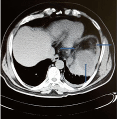
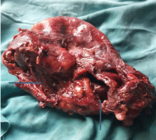
The CT- scan of the abdomen revealed splenomegaly (17 X 8 cm), suspicious splenic infarction and suspicious splenic abscess in progression (Figure 1). After the admission, we have started to treat a patient by imipenem- cilastatine, a broad-spectrum antibiotic. Three days after admission patient underwent surgery with the aim to remove the septic source and the diseased organ. About 90% of spleen parenchyma was destructed due to abscess. Spleen was like bag filled with pus, and an open splenectomy was performed (Figure 2). From splenic abscess has been isolated enterococcus faecalis. The patient was discharged on the seventh postoperative day. Fourteen days after splenectomy, a patient has been vaccinated by pneumococcal, meningococcal, and Haemophylus Influence (Hib) vaccines. A month after splenectomy patient was in very good condition.

    Figure 1: CT showing hypodense fluid collection in the upper and the middle
part of the spleen, and hypodense the rest part of the spleen.

Figure 2: Parenchymal destruction of the spleen due to abscess.
Splenic abscess is a rare entity, with high mortality rate, up to 47%. The most common causes of splenic abscesses are haematogenous spread originating from an infective focus elsewhere in the body, urinary tract infection, infective endocarditis, pneumonias, pelvic infections etc [1,2]. Splenic infarction resulting from systemic disorders such as hemoglobinopathies (especially sickle cell disease), leukemia, polycythemia, or vasculitis, can become infected and evolve into splenic abscess [3,4]. Splenic abscess can be treated by percutaneous drainage or surgical interventions (open or laparoscopic splenectomy) [5,6].
References
- Bayer AS, Bolger AF, Taubert KA, Wilson W, Steckelberg J, Karchmer AW, et al. Diagnosis and management of infective endocarditis and its complications. Circulation. 1998; 98: 2936-2948.
- Smyrniotis V, Kehagias D, Voras D, Fotopoulos A, Lambrou A, Kostopanagiotou G, et al. Splenic abscess. An old disease with new interest. Dig Surg. 2000; 17: 354-357.
- Tung CC, Chen FC, Lo CJ. Splenic abscess: an easily overlooked disease?. Am Surg. 2006; 72: 322-325.
- Al-Salem AH. Splenic complications of sickle cell anemia and the role of splenectomy. ISRN Hematol. 2011. 2011: 864257.
- Ooi LL, Leong SS. Splenic abscess from 1987 to 1995. Am J Surg. 1997; 174: 87-93.
- Carbonell AM, Kercher KW, Metthews BD, Joels CS, Sing RF, Heniford BT. Laparoscopic splenectomy for splenic abscess. SurgLaparoscEndoscPercutan Tech. 2004; 14: 289-291.
