Abstract
Non-occlusive mesenteric ischemia is frequent in critically ill patients with hemodynamic instability. Plasma Lactate is a marker of tissue hypoperfusion and its diagnostic significance in the setting of mesenteric ischemia remains controversial. We present a case of severe non-occlusive mesenteric ischemia without lactate elevation over normal values although extended intestinal necrosis requiring right hemicolectomy and total ileoectomy was present. Serial lactate concentration measurements were insufficient to detect intestinal hypoperfusion and predict the length of bowel necrosis.The pre-and postoperative clinical monitoring was subsequently guided by vasoactive medication demand.
Keywords: Non-occlusive mesenteric ischemia; Intestinal necrosis; Lactate; Lactate acidosis
Introduction
Acute Mesenteric Ischemia (AMI) is a life-threatening reduction of the intestinal perfusion observed after arterial or venous occlusion or non-occlusive arterial infarction secondary to vasospasm more often in critically ill patients with hemodynamic instability under vasoconstrictive medication. Plasma lactate has been tested as a marker of mortality [1] and Irreversible Transmural Intestinal Necrosis (ITIN) [2] in the setting of acute mesenteric ischemia. Although lactate concentration several hours before death or surgical interventionwas recognized as independent risk factor predicting the severity of AMI [1-3] its role in the diagnosis of this condition in early stageremains controversial. Lactate is an unspecific marker of organ hypoperfusion and becomes elevated only after significant tissue damage [4]. Plasma D-lactate was initially introduced as a more specific alternative to routinely measured stereoisomer L-lactate in the sense of its anaerobic bacterial origin [5]. However experimental studies put the significance of D-lactate under question [6] and novel markers of gut mucosal damage like intestinal fatty acid-binding protein were recommended [4,7]. According to many observations lactate remains a reliable marker of mesenteric ischemia detection secondary to acute aortic dissection [8,9]. We present a case of a severe non-occlusive mesenteric ischemia with gut necrosis requiring right hemicolectomy and total ileoectomywithout lactate elevation over normal values.
Case Presentation
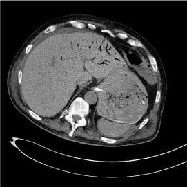
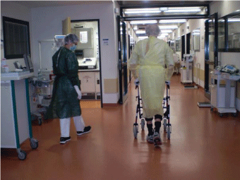
The 76-year-old man with unremarkable medical history was treated in our intensive care unit with Acute Respiratory Distress Syndrom (ARDS) by eosinophilic pneumonia, occurred without a detectable pathogen and by missing underlying immunologic insufficiency. During the clinical course hypoxemia secondary to lung failure ensued and support of Veno Venous Extra Corporeal Membrane Oxygenation (VV ECMO) was required. The patient received treatment with steroids and broad spectrum antibiotics. Having recovered, VV ECMO was removed and weaning from sedation was initiatedunder mechanical ventilation. 48 hours after sedative withdrawal, respiratory effort was observed and assisted ventilation was started. A sinus tachycardia (110-120 beats/min) and elevated body temperature (37.5-37.8°C) were evaluated as cardiovascular reaction during the recovery phase. 12 hours later acute arterial hypotension occurred, requiring vasoconstriction with Nor-adrenalin, as application of crystalloids was insufficient. Because of the simultaneous appearance of respiratoryinstability with progressive hypoxemia, a new sepsis course with lung focus as a result of the weaning phase was supposed. Considering the Central Lineassociated Bloodstream Infection all lines and catheters were changed. Echocardiography excluded cardiogenic shock. Physical examination revealed meteorismus with hypoactive abdominal sounds but abdominal Ultrasonography was at the time point unremarkable. We initiated osmotic and stimulant laxatives as by intensive careassociated constipation and decided to wait and observe. 4 hours later abdominal circumference increased manifestly and abdominal Ultrasonography revealed aerobilia, which was a peculiar finding, as the patient had no history of a biliary system intervention. Because of the severe clinical deterioration, we decided to perform a contrastenhanced whole-body computed tomography in order to expand our diagnostic panel. The computed angiography excluded pulmonary embolism as a cause ofrespiratory and cardiovascular instability. An extended acute mesenteric ischemia occurring in the blood supply region of the superior mesenteric artery with hepatic portal venous gas and pneumatosisintestinaliswas diagnosed (Figure 1) and the patient underwent an emergency laparotomy with right hemicolectomy and ileoectomy because of irreversible bowel necrosis. An atherosclerotic disease or thrombotic occlusion of the abdominal aorta and its mesenteric branches was not present and we presumed an acute nonocclusive mesenteric ischemia due to vasospasmus. The postoperative course was unremarkable and clinical recovery was achieved eight weeks later (Figure 2).

    Figure 1: An extended non-occlusive mesenteric ischemia with hepatic portal
venous gas and pneumatosis intestinalis was diagnosed.

    Figure 2: Clinical recovery eight weeks after severe non-occlusive mesenteric
ischemia treated with right hemicolectomy and ileoectomy.
Discussion
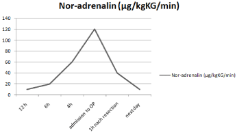
In our case a severe non-occlusive mesenteric ischemia was occurred under the use of vasoactive medication, which was implemented in order to treat septic shock. We believe that the septic shock was of pulmonary origin and ischemia presented secondarily. However the pathophysiological cascade of our case remains unclear. For example, non-occlusive mesenteric ischemia could have happened primarily to pulmonary and hemodynamic deterioration. An argument to support this theory could be the immediate recovery of vasoconstrictive drugs right after ischemic bowel resection (Figure 3). Experimental animal models have registered the appearance of multiple organ failure and Acute Respiratory Distress Syndrome (ARDS) as a result of mesenteric ischemia [10, 11]. This can be explained through the inflammatory cascade with cytokines production. Nevertheless, multiple organ failure in our case was partly responsible for the delayed diagnosis, because of the missing focus.

    Figure 3: Pre-and postoperative monitoring of mesenteric hypoperfusion was
performed with assistance of catecholamine demand.
The second factor was the absence of lactate elevation. Lactate values remained normal throughout the clinical course, although extensive mesenteric ischemia with hepatic portal venous gas and pneumatosisintestinalishas occurred. The portal venous gas explains the ultrasonographic detection of air in the liver. We believe that systemic lactate was normal because of its sufficient hepatic elimination. Studies have shown that lactate hepatic uptake increases in response to elevated splanchnic production and development of arterial hyperlactatemia can be prevented if systemic hypovolemiais treated by fluid resuscitation during mesenteric hypoperfusion [12].
Our clinical observation in this case consists with experimental findings, where higher lactate values can predict mortality [1,13] but serial lactate concentration measurements are insufficient to predict the length of intestinalnecrosis [13]. However, other markers of mesenteric ischemia like stereoisomer L-lactate and intestinal fatty acid-binding protein were not tested in our case. Because of the absent lactate elevation, pre-and postoperative monitoring of mesenteric hypoperfusion was performed with assistance of catecholamine extension. Figure 3 presents the progressive demand of vasoactive medication beginning 12h before surgery and rapidly dropping after resection of necrotic bowel. The extension of bowel resection was initially defined according to intra-operative findings.
Finally, we believe that postoperative clinical course was uneventful and full recovery was achieved probably because of missing mesenteric atherosclerosis, which could have induced a secondary mesenteric occlusion [14].
References
- Leone M, Bechis C, Baumstarck K, Ouattara A, Collange O, Auqustin P, et al. Outcome of acute mesenteric ischemia in the intensive care unit: a retrospective, multicenter study of 780 cases. Intensive Care Med. 2015; 41: 667-676.
- Nuzzo A, Maggiori L, Ronot M, Becq A, Plessier A, Gault N, et al. Predictive Factors of Intestinal Necrosis in Acute Mesenteric Ischemia: Prospective Study from an Intestinal Stroke Center. Am J Gastroenterol. 2017; 112: 597- 605.
- Studer P, Vaucher A, Candinas D, Schnuriger B. The value of serial serum lactate measurements in predicting the extent of ischemic bowel and outcome of patients suffering acute mesenteric ischemia. J Gastrointest Surg. 2015; 19: 751-755.
- Demir IE, Ceyhan GO, Friess H. Beyond lactate: is there a role for serum lactate measurement in diagnosing acute mesenteric ischemia? Dig Surg. 2012; 29: 226-235.
- Yao YM, Yu Y, Wu Y, Lu LR, Sheng ZY. Plasma D (-)-lactate as a new marker for diagnosis of acute intestinal injury following ischemia-reperfusion. World J Gastroenterol. 1997; 3: 225-227.
- Nielsen C, Mortensen FV, Erlandsen EJ, Lindholt JS. L- and D-lactate as biomarkers of arterial-induced intestinal ischemia: an experimental study in pigs. Int J Surg. 2012; 10: 296-300.
- Salim SY, Young PY, Churchill TA, Khadaroo RG. Urine intestinal fatty acidbinding protein predicts acute mesenteric ischemia in patients. J Surg Res. 2017; 209: 258-265.
- Kurimoto Y, Morishita K, Fukada J, Kawaharada N, Komatsu K, Yama N, et al. A simple but useful method of screening for mesenteric ischemia secondary to acute aortic dissection. Surgery. 2004; 136: 42-46.
- Morisaki A, Kato Y, Motoki M, Takahashi Y, Nishimura S, Shibata T. Delayed Intestinal Ischemia after Surgery for Type A Acute Aortic Dissection. Ann Vasc Dis. 2015; 8: 255-257.
- Steinberg J, Halter J, Schiller H, Gatto L, Nieman G. The development of acute respiratory distress syndrome after gut ischemia/reperfusion injury followed by fecal peritonitis in pigs: a clinically relevant model. Shock. 2005; 23: 129-137.
- Collange O, Charles AL, Lavaux T, Noll E, Bouitbir J, Zoll J, et al. Compartmentalization of Inflammatory Response Following Gut Ischemia Reperfusion. Eur J VascEndovasc Surg. 2015; 49: 60-65.
- Jakob SM, Merasto-Minkkinen M, Tenhunen JJ, Heino A, Alhava E, Takala J. Prevention of systemic hyperlactatemia during splanchnic ischemia. Shock. 2000; 14: 123-127.
- Studer P, Vaucher A, Candinas D, Schnuriger B. The value of serial serum lactate measurements in predicting the extent of ischemic bowel and outcome of patients suffering acute mesenteric ischemia. J Gastrointest Surg. 2015; 19: 751-755.
- Reissfelder C, Sweiti H, Antolovic D, Rahbari NN, Hofer S, Buchler MW, et al. Ischemic colitis: who will survive? Surgery. 2011; 149: 585-592.
