Clinical Image
Austin Emerg Med. 2015; 1(1): 1005.
Diaphragmatic Hernia Resembling Tension Pneumothorax
Luis Angel MA* and Collazos SS
Department of General Surgery, Universidad de Quintana Roo, Mexico
*Corresponding author: Luis Angel Medina Andrade, Department of General Surgery, Universidad de Quintana Roo, Mexican Social Security Institute (IMSS), Cancún, Quintana Roo, Mexico
Received: December 04, 2015; Accepted: December 07, 2015; Published: December 09, 2015
Clinical Image
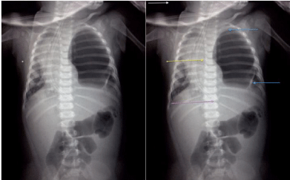
An 8-months male patient presents at Emergency Department with respiratory distress. His parents refer beginning of symptoms two days ago with worsening 10 hours previous to first contact. After initial minutes of initial evaluation and x-ray performance the patient present cardiorespiratory failure and cardio pulmonary reanimation was given to him. At physical exam without ventilation in left hemithorax. A hematoma in the left axillary region was noted and a tension Pneumothorax suspected. After the first two cycles of compressions surgery service was informed of the case. By the mentioned facts and a radiograph resembling a tension Pneumothorax with displacement of vessels and hearth to the right hemithorax, a decompression punction was performed in medial clavicular line second intercostal space obtaining air and a white fluid resembling milk. A diaphragmatic hernia was suspected in this moment but pulse was not obtained after reanimation maneuvers and exploratory laparotomy could not be performed.

    Figure 1:  White arrow: Top of the image.
Yellow arrow: Great vessels and hearth displacement.
Purple arrow: End of nasogastric tube.
Blue arrows: Gastric wall.