Short Communication
Austin Emerg Med. 2016; 2(1): 1006.
Urgent Surgery for Severe Infective Endocarditis
Xuhe Gong and Guogan Wang*
State Key Laboratory of Cardiovascular Disease, Fuwai Hospital, National Center for Cardiovascular Diseases, Chinese Academy of Medical Sciences and Peking Union Medical College, Beijing, People’s Republic of China
*Corresponding author: Guogan Wang, State Key Laboratory of Cardiovascular Disease, Fuwai Hospital, National Center for Cardiovascular Diseases, Chinese Academy of Medical Sciences and Peking Union Medical College, Beijing, 100037, People’s Republic of China
Received: December 03, 2015; Accepted: January 05, 2016; Published: January 12, 2016
Abstract
Background: The critical role of urgent surgery for severe Infective Endocarditis (IE) has been well established. Here, we want to highlight the importance of urgent surgery for severe IE to prevent serious consequences.
Methods: We present a report of severe aortic and mitral valve IE in a 47-year-old man. Echocardiography revealed large, mobile mitral valve vegetation with severe aortic and mitral regurgitation. Patient presented with persistent fever together with pulmonary symptoms and heart failure, and was diagnosed with infective endocarditis. The treatment was challenging due to multiple serious complications, which made the drug treatment poor. After two weeks, when his temperature dropped and stayed down for two days, a surgical intervention was done. During the postoperative period, multiple cerebral infarctions developed in the patient.
Result: The treatment was completed in seven weeks with full recovery.
Conclusion: Infective endocarditis may present with various clinical situations that may be life-threatening, urgent surgery is necessary in severe infective endocarditis.
Keywords: Infective endocarditis; Urgent surgery; Pneumonia
Introduction
The demography and microbiology of IE have changed in recent decades, although risk factors such as rheumatic heart disease have become less prevalent, intravenous drug use, degenerative valvular disease, and health care-associated infection are more common. The incidence of IE has also remained constant over time and affects 5-15 per 100,000 people per year; it remains as a devastating condition.
Case Report
A 47-year-old male was brought to the emergency department with severe dyspnea. The initial symptom, fever, was present two months. He was febrile with exertional dyspnea and observed in a local hospital two weeks ago. X-ray showed signs of bilateral pulmonary infection, echocardiography demonstrated vegetation on the mitral valve. Streptococcus viridans and Enterococcus faecalis was isolated in blood cultures. The condition deteriorated with anemia, orthopnea and fatigue, so he was transferred to our hospital.
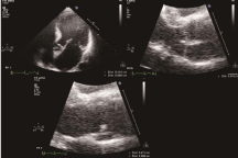
On presentation, the patient had a BP of 123/59mmHg, HR of 96bpm, temperature of 36.5oC. Pertinent laboratory values were: WBC 14.6*109/L, ALT 96IU/L, Albumin 28g/L, Chest radiograph showed bilateral alveolar opacities, Cardiac auscultation revealed a 3/6 grade systolic murmur radiating to the axilla, Coarse bibasilar rales were heard on chest auscultation. The bedside ultrasonography was immediately performed, which revealed large, mobile vegetation (maximum 4×6mm) on the mitral anterior lobe, mitral valve prolapse and severe aortic regurgitation (Figure 1).

    Figure 1:  Echocardiography demonstrated large, mobile vegetations
(maximum 6 × 4 mm) on the mitral anterior lobe, mitral valve prolapse.
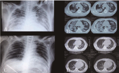
Penicillin 24,000,000IU/day and amikacin 0.2g/day was initiated, the serial blood cultures were all negative, sputum culture was positive for Pseudomonas aeruginosa and Klebsiella pneumoniae. On the third day, echocardiography revealed vegetation on the non- coronary aortic valve with fistula and severe regurgitation. Since white blood cells continued to rise, the treatment was replaced with linezolid 0.6g/day + moxifloxacin 0.4g/day, but fever continued and no reduction in the size of the vegetation was observed, X-ray and CT showed a lot of exudative inflammation (Figure 2). His condition deteriorated with hepatic insufficiency, the final treatment was replaced with meropenem 1g q8h and teicoplanin 0.3g/day. On the day 15, the temperature returned to normal, two days later, The patient was performed an aortic and mitral valve replacement surgery, two separate 1- and 1.2-cm perforation were found on the anterior leaflet of mitral valve and non-coronary valve, respectively, to which the vegetation were adhered.

    Figure 2:  X-ray and CT of patient.
A and B: the chest X-ray and CT before operation.
C and D: the chest X-ray and CT after operation.
Further conservative antibacterial and anti-heart-failure therapy were administered, pulmonary edema were absorbed gradually (Figure 2). On the 12th day of post-operation, combined aphasia and incontinence of feces and urine appeared. The brain CT showed multiple infarcts, measures to reduce intracranial pressure and warfarin was administered. After half a year, the brain CT showed complete remission.
One month after operation, echocardiography confirmed no vegetation and the mechanical valve worked well. The patient resumed normal activities without any mishaps during the 3 years’ follow-up.
Discussion
The demography and microbiology of IE have changed in recent decades, Although risk factors such as rheumatic heart disease have become less prevalent, intravenous drug use, degenerative valvular disease are more common [1], it remains as a devastating condition. Heart failure and infection are the relatively common and lifethreatening complications, which is involved in this patient. What’s more, splenic infarction, cerebral infarction, drug allergies and diarrhea developed in the patient. This confirms IE may present with various clinical situations.
Despite appropriate antimicrobial and anti-heart-failure therapy, which is recommended, there was no improvement of his endocarditis. But if untreated, it would pose a potential life-threatening situation, so the surgery is necessary, which has also been recommended in the European Society of Cardiology 2009 guidelines on the prevention, diagnosis, and treatment of infective endocarditis [2].
In the guidelines, a reduction in antibiotic prophylaxis and a recommendation to operate early on patients with large vegetations are the two main changes [3]. The surgical intervention is a potential lifesaving procedure, which is required in 50% of cases [4]. Indications for early surgery include persistent fever despite adequate antibiotic treatment, refractory heart failure and thromboembolic events, the first two indications present in the patient. The combination of aggressive antibiotic administration and early surgical intervention is lifesaving.
Conclusion
IE is a systemic and changing disorder, Patients today are much older and have more comorbidities, which is different from the past decade, mortality remains very high. With the increasing evidence supporting urgent surgical intervention in IE [5], we should recognize that performing a technically sound operation at the proper time are keys to optimizing outcomes; Surgery delayed may become surgery denied.
References
- Evans CF, Gammie JS. Surgical management of mitral valve infective endocarditis. Semin Thorac Cardiovasc Surg. 2011; 23: 232-240.
- Harrison JL, Prendergast BD, Habib G. The European society of cardiology 2009 guidelines on the prevention, diagnosis, and treatment of infective endocarditis: key messages for clinical practice. Pol Arch Med Wewn. 2009; 119: 773-776.
- Taylor J. The 2009 ESC Guidelines for management of infective endocarditis reviewed. Eur Heart J. 2009; 30: 2185-2194.
- Baig W, Sandoe J. Infective endocarditis. Clin Med. 2010; 10: 188-191.
- Kang DH, Kim YJ, Kim SH, Sun BJ, Kim DH, Yun SC, et al. Early surgery versus conventional treatment for infective endocarditis. N Engl J Med. 2012; 366: 2466-2473.