Editorial
Austin Emerg Med. 2016; 2(1): 1007.
The Emerging “Echoscopic” Paradigm
Favier JC*
Scorff Hospital, Anesthesia Service Lorient, France
*Corresponding author: Favier JC, Scorff Hospital, Anesthesia Service Lorient, France
Received: December 09, 2015; Accepted: January 05, 2016; Published: January 12, 2016
Editorial
Ultrasound manufactures propose us until last year’s very small US devices and each generation is cheaper and more powerful.
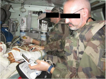
These devices are a real revolution leading to shifts in our way of working and thinking (Figure 1). It is already possible to use them in place it was impossible to use ultrasound before. It was therefore possible to use US in the field, during evacuation in the ambulance, helicopter for example.

Figure 1: In the field, during evacuation in the ambulance, helicopter.
When these devices appeared, opinion leaders had to consider the situations where “echoscopy” would be useful. Of course, prehospital location is interesting and a widespread use is seen in particular in Europe.
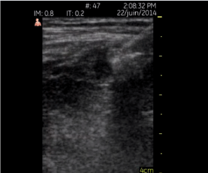
But the further improvements proposed by manufacturers: multiprobe machines, wider screens lead us to thing that these devices are not only echoscopes. Senior ultrasound physician can use them in many other situations for example during echoscopic guidance for neural blockade (Figure 2), pleural effusion treatment, line placement. And it is possible to put the whole device in a sterile sock to perform real and accurate guidance. The distance between the target and the screen is small so there are poor ergonomic difficulties to organize your professional area of work even if the picture is small.

Figure 2: Femoral “echoscopic” guided block.
As a consequence, it induced in my teaching method to promote better ergonomic installation with the standard and compact ultrasound machines. This collateral consequence of this observation. Ergonomy is clearly a point under teached until now, inducing regularly difficulties during a procedure and back and neck pain for the physician.
It is sometimes much more easy to learn how to use US devices if the first US device used is one of these small machines because of intuitive ergonomic positioning. Teaching cardiac US examination to US beginners with a small device I was stunned by the way beginners used US intuitively choosing the right ergonomy in contrast with the path I personally had to drive to improve my non ergonomic position when I was a resident, learning the technique.
Furthermore it induces also a different view of what will be the right place in actual and further medical practice
So hygiene and ergonomy can be improved but for some points in particular echoguidance there is a prealable conditions to have a large improvement in our practices induced by the small devices. It is necessary to be a good US performer. And probably it is necessary to learn ultrasound standards on compact or normal machines before wide small device use to reach a minimal level in ultrasound diagnostic and guidance.
If 10 years ago the small US devices could be possibly considered as new devices facilitating ultrasound use in beginners. Actually this positioning could always be argued but it is possible to also have now a diametrally opposite point of view as a specific useful device for senior confirmed users.
In the early 2010 I personally didn’t believe in such machines but I was wrong.
As an ultrasound teacher I actually have a very different point of view. Small and big US machines are complementary. And it is still and obviously necessary to better teach ergonomy.
Other paths of use are going to emerge in particular because of increasing number of obese patients and drug users to manage (line placement at the bedside), leading to spare time in patient management. Lack of anesthesiology physician in France leads to extensive backup anesthesiologist employment in hospitals and in the private institutions. If you have such a device it is reassuring to go in a new institution and discover a new environment. This was my case before choosing the actual institution i work in.
In the operative room it is possible to use the small devices limiting transoesophagal exam by example using the transhepatic view of the heart and the inferior cava vein to guide preload management [1]. This is possible without the necessity to interrupt the surgical procedure. It is not possible to make easily the exam with a standard and even a compact machine because the surgeon has to remove to let you make the exam. We also underuse the jugular and superior cava vein diameter variations that we can integrate in our medical approach with small device US machines and also with standard US machines of course.
So the small devices came about ten years ago and many of us didn’t know how to use them. Now it is possible to discuss what becomes to be feasible.
Of course there will be many other paths of use that I still ignore. And if the manufacturers propose us to connect the small machines to wide screen. What are we going to be able to do?? Manufactures foregoing our needs?
References