Case Report
Austin Emerg Med. 2016; 2(1): 1008.
Ureteropelvic Junction Obstruction in Adult Patient with Abdominal Mass
Farzad Rahmani¹*, Asghar Jafari Roohi², Arezoo Nejabatian² and Hanieh Ebrahimi Bakhtavar¹
¹Road Traffic Injury Research Center, Tabriz University of Medical Sciences, Iran
²Emergency Medicine Research Team, Tabriz University of Medical Sciences, Iran
*Corresponding author: Farzad Rahmani, Emergency Medicine Department, Sina Hospital, Azadi Avenue, Tabriz University of Medical Sciences, Tabriz, Iran
Received: November 18, 2015; Accepted: December 10, 2015; Published: January 12, 2016
Abstract
Patients in the different ages may refer to the Emergency Department (ED) with complaining of the abdominal mass. Based on history, clinical examination, the patient’s age and the location of mass, several differential diagnoses are discussed. In this report, we represent a 23-year old female patient which had referred to our ED with complaining about the pain and swelling of the right side of the abdomen. After taking the necessary measurements, Ureteropelvic Junction Obstruction (UPJO) diagnosis was made for patient. The renal tissue degradation was happened as a consequence of delayed diagnosis. The patient was underwent nephrectomy. Since UPJO is usually seen in children, we decided to introduce this patient as a case report. Emergency physicians must constantly think about UPJO in patients with abdominal mass, so that they can save the function of kidney with accurate and rapid diagnosis.
Keywords: Abdominal pain; Ureteral obstruction; Nephrectomy
Introduction
Abdominal masses are one of the causes for which patients come to the emergency department. Abdominal masses have many differential diagnosis including life-threatening ones such as abdominal aortic aneurysm to cases with less risk. The differential diagnosis includes tumors, ovarian cysts, inflammatory bowel disease, gastrointestinal tumors, abdominal hernia, kidney tumors, kidney hydronephrosis, lymphoma and pancreatic cysts [1]. Abdomen is divided anatomically into four quadrants. According to the location of a mass; the specific differential diagnosis is discussed. In a patient who refers with an abdominal mass, after conducting the examination, it is required to conduct laboratory tests and imaging in order to diagnose the origin and the probable diagnosis of mass [2]. Here, we report a 23-year old female patient which had referred to our ED with complaining about the pain and swelling of the right side of the abdomen. After gathering the necessary measurement, UPJO diagnosis was made for patient.
Case Presentation
The presented patient was 23-year old female which had referred to our ED with complaining about the pain and swelling of right flank and right upper quadrant of the abdomen. The patient told us she had the intermittent abdominal pain without any relation to feeding, movement and changing position since about one and a half year ago, pain has been more in right side of abdomen with radiating to the lower portions. The pain was ambiguous and was subsided spontaneously. The severity and duration of her pain has increased gradually since a month ago before referring to our ED, quality of the pain has become constantly and swelling of the right flank also gradually has been added to the patient’s symptoms. Because of the patient’s pain was intensifying and intolerable from a day ago, she referred to ED. During urination, she felt a severe pain and nausea. Also, she had urinary frequency. Her bowel habit was normal. There was a history of urinary symptoms and recurrent urinary infection in this patient but the complete treatment and the clinical examination and laboratory and imaging findings had not been conducted. She was married and her sexual history had not any significant problem. She used antibiotics for urinary infection. She had not any trauma before. There wasn’t any other previous medical history and medication. The vital signs were as follows:
BP: 110/70, PR: 92/min, RR: 14/min, Spo2: 97% (in room air), BT: 36.7 ‘C (axillary).
The patient was alert and oriented in examination. Examination of the heart and lungs were normal. In abdominal examination, the swelling and a palpable 10 cm mass was in Right Upper Quadrant (RUQ) and also progressed to the Right Lower Quadrant (RLQ). The mass was non pulsatile and firm in palpation. She had tenderness in right flank and right costovertebral angle. Examinations of other areas hadn’t any problem. The results of the blood tests are seen in Table 1. There wasn’t any pathological finding in urine analysis.
    Element
    Result
    Normal range


    WBC
    12*103/mm3
    (4-10)* 103/mm3


    Hb
      Hct
    8.1 gr/dl
      25%
    12-16 gr/dl
      36-46%


    FBS
    95 mg/dl
    70-115 mg/dl


    Urea
    26 mg/dl
    15-40 mg/dl


    Cr
    0.9 mg/dl
    0.7-1.4 mg/dl


    Amylase
    65 IU/L
    ˂100 IU/L


    Sodium
    139 mEq/L
    135 - 145 mEq/L


    Potassium
    4.1 mEq/L
    3.5-5.0 mEq/L


    Ca++
    0.10 mmol/L
    1.1-1.35 mmol/L


    ALT
    12 IU/L
    7-56 IU/L


    AST
    16 IU/L
    10-40 IU/L


    Alk. P
    83 IU/L
    44 to 147 IU/L


    Beta HCG
    ˂10 unit


    HCV Ab
    Negative


    HBs Ag
    Negative
Table 1: Laboratory Findings of Patients.
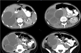
We carried out emergency abdominal Ultrasonography (US) for patient, and US was showed a mass with several cysts in RUQ that the right kidney was not seen naturally in its place, we could not printed of patient’s US images. Differentiation of the mass and the origin of mass were not clear with US. Then we requested abdominal and pelvis CT scans for a more accurate diagnosis. As it is seen in Figure 1, the right kidney was observed with severe hydronephrosis so that the normal kidney tissue has been destroyed. The patient with a probability diagnosis of UPJO was admitted in the surgical ward and then the patient was undergone the abdominal surgery and nephrectomy. After that, the patient has been discharged from surgery ward after one week without any problem.

    Figure 1:  Abdominal CT scan of patient. This image shows a polycystic mass
at abdominal right side.
Discussion
One of the causes of abdominal masses is hydronephrosis of kidney. Hydronephrosis means the dilation of renal calyces and is caused by obstruction in the urinary output path from the kidney. UPJO is one of the causes of hydronephrosis which can be presented as abdominal masses more commonly in children and neonates than in adults. Treatment of hydronephrosis is very important because it increases the risk of kidney dysfunction, infection, stone and pain [3,4]. There is limited information about the UPJO in adults [5], Because of increasing the quality of screening methods in neonatal and even prenatal periods, the cases of UPJO in adults has become rare [6]. Unlike children in whom all hydronephrosis cases do not require the intervention, in adults, the treatment of UPJO is necessary in order to improve the renal function and prevent renal damage [7]. An obstruction in UPJ in the congenital cases is an anatomical defect caused by impairment in the intrauterine evolution, while UPJO in adults and the acquired cases can be result of the upper urinary tracts infection, stone and trauma. UPJO emerges in adults with an acute colicky pain, flank chronic pain and hematuria in few cases, urinary tract infections with or without pyelonephritis, but the abdominal distension because of the presence of mass is a rare symptom [8,9]. One of the causes of UPJO can be a crossing vessel that makes pressure on ureter and leads to the obstruction [10].
The abdominal CT scan of introduced patient showed the severe hydronephrosis at right kidney with evidences of the renal tissue degradation so that all of the kidney tissue was filled by the severe and extensive hydronephrosis and it led to palpable mass in RUQ and right flank. The patient had referred with the abdominal mass symptoms. It is remarkable that hydronephrosis with this severity and UPJO as the cause is an uncommon and rare in adults. Hydronephrosis is a result of urinary tract obstruction which is caused by multiple etiologies, but during operation, surgeon did not disclosure any other problems such as aortic aneurysm, renal stone, crossed vessel and etc. Hydronephrosis is also more common in males than females while the introduced patient was female. Evaluation and diagnosis of hydronephrosis are conducted by Sonography, CT scan and venous Pyelography [11].
In this patient because of her presentation with abdominal mass and unknown origin of the mass, Sonography was conducted at first but it was unable to differentiate between the hepatic and renal origin of mass, for this reason, CT scan of abdomen and pelvis with Contrast was conducted. CT scan showed the severe hydronephrosis of kidney and its parenchymal degradation with the possibility of UPJO. So that, the patient underwent an abdominal surgery to remove right kidney. During surgery, given the fact that all of right kidney was cystic, and normal kidney tissue was not observed, nephrectomy was done. If the UPJO was diagnosed in early stage, probably these problems were not occurred.
Renal severe hydronephrosis is one of the causes of abdominal masses. UPJO is one of the diseases that cause hydronephrosis. This disease although is more common in children but its incidence is not unexpected in adults and we must constantly think about this diagnosis so that we can save the function of kidney with accurate and rapid diagnosis.
Acknowledgment
We acknowledge all staffs of emergency department and general surgery ward of Sina Hospital, Tabriz, Iran.
References
- Rahhal RM, Eddine AC, Bishop WP. A Child with an Abdominal Mass. Pediatric Rounds. 2006; 37-42.
- Calinescu C, Jackson I, Mauriello M, Chern I, Schwarz ER. A Systematic Approach for the Assessment and Diagnosis of Abdominal Pain in the Premenopausal Female. American Journal of Clinical Medicine. 2011; 8: 160-163.
- Nicholas J Hellenthal, Sasha A Thomas, Roger K Low. Rapid onset renal deterioration in an adult with silent Ureteropelvic junction obstruction. Indian Journal of urology. 2009; 25:132-133.
- Laurence S Baskin. Congenital Ureteropelvic junction obstruction. Melanie S Kim (editor), Up-to-date. 2015.
- Hartman GE, Shochat SJ. Abdominal mass lesions in the newborn: diagnosis and treatment. Clin Perinatol. 1989; 16: 123-135.
- Capello SA, Kogan BA, Giorgi LJ, Kaufman RP. Prenatal ultrasound has led to earlier detection and repair of ureteropelvic junction obstruction. J Urol. 2005; 174: 1425-1428.
- Park JM, Bloom DA. The pathophysiology of UPJ obstruction. Current concepts. Urol Clin North Am. 1998; 25: 161-169.
- Braga LH, Liard A, Bachy B, Mitrofanoff P. Ureteropelvic junction obstruction in children: two variants of the same congenital anomaly? Int Braz J Urol. 2003; 29: 528-534.
- Gnanapragasam VJ, Armitage TG. Laparoscopic pyeloplasty, initial experience in the management of UPJO. Ann R Coll Surg Engl. 2001; 83: 347-352.
- Conlin MJ. Results of selective management of ureteropelvic junction obstruction. J Endourol. 2002; 16: 233-236.
- Grasso M, Caruso RP, Phillips CK. UPJ Obstruction in the Adult Population: Are Crossing Vessels Significant? Rev Urol. 2001; 3: 42-51.