
Case Report
Austin Emerg Med. 2016; 2(4): 1025.
ECG Lead Misplacement -Fool Me Once Shame on You, Fool Me Twice Shame on Me
Grossi I1 and Lynch RM2*
¹Senior House Officer in Emergency Medicine, Midland Regional Hospital, Mullingar, Co. Westmeath, Ireland
²Consultant in Emergency Medicine, Midland Regional Hospital, Mullingar, Co. Westmeath, Ireland
*Corresponding author: Richard M Lynch, Consultant in Emergency Medicine, Midland Regional Hospital, Mullingar, Co. Westmeath, Ireland
Received: March 03, 2016; Accepted: April 18, 2016; Published: April 20, 2016
Abstract
ECG lead misplacement can dramatically alter the appearance of the ECG. If this is not detected significant and profound errors in treatment can ensue. We report the case of a 42 year old male who presented following a fall down a flight of stairs. An ECG was performed and this was compared with a previously recorded ECG which was filed in the patient’s hospital record. Two separate limb lead misplacements were present on this ECG even though classical features of both limb lead misplacements were present. Not only was this ECG filed in the in-patient records the ECG was not signed off by a senior doctor.
Highlights: ECG lead misplacements alter the ECG diagnosis in approximately 25% of cases.
Use of the “REVERSE mnemonic” has the potential to highlight incorrect lead placement.
A near-flat line in lead II is characteristic of right arm-right leg misplacement.
In RA-LA misplacement P-QRS-T complexes are negative in lead I and positive in a VR.
Keywords: ECG lead misplacement; “REVERSE” mnemonic; Right arm -right leg misplacement; Right arm- left arm
Introduction
The Electrocardiogram (ECG) is one of the most important and most frequently requested investigations in the management of the acutely unwell patient [1]. It is inexpensive, quick and easy to record and this record is easy to reproduce if appropriate attention to the placement of electrodes is given [2]. In spite of this lead misplacements do occur which, if not recognized, may have disastrous consequences with patients receiving the wrong treatment or not being treated at all based on the features of an incorrectly acquired ECG [3].
We describe a previously unreported case in which two separate limb lead misplacements occurred simultaneously on the same ECG even though the characteristic features of both were clearly evident. A review of the ECG appearances of both examples of misplacement is presented together with tips on how to recognize these lead misplacements.
Case Presentation
A 42 year old male, with a background history of epilepsy, presented to our Emergency Department (ED) after falling down a full flight of stairs. He stated that he felt like he was going to have a seizure while at the top of the stairs. The next thing that he remembered was waking up at the foot of the stairs. He complained of injuries to his head and neck.
On examination vital signs were within normal limits. Tenderness was elicited over the occiput and the upper cervical spine. A CT scan of his head and neck was performed, both of which were normal.
An ECG was performed to determine if his fall might have been due to an arrhythmia. This was normal (Figure 1). Due to the potential causes and consequences of the fall the in-patient records were retrieved to search for any relevant past medical conditions and also to compare his ECG with previously recorded ones. One of the ECGs in the in-patient records, which had been recorded one year earlier, revealed classical features of not one but two different types of ECG limb lead misplacement, namely right arm – right leg misplacement and right arm – left arm misplacement (Figure 2). Of note this abnormal ECG does not appear to have been “signed off” by a senior doctor. It was filed in the in-patient notes and even though previously recorded ECGs with no evidence of lead misplacement were available for comparison these two limb lead misplacements went undetected. It was not possible to identify who had missed the lead misplacements. It was never our intention to reprimand any member of staff as a result of these errors. We chose to educate all medical staff and nursing staff, regardless of status, on the appearance of limb lead misplacements. We hope that this intervention will help reduce the overall numbers of lead misplacements, and in particular those which heretofore have gone undetected.
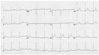
Figure 1: Normal sinus rhythm.
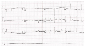
    Figure 2:  ECG with characteristic features of both right arm-right legand right
arm-left arm misplacement.
Discussion
ECG lead misplacements alter the diagnosis in approximately 25% of cases [4] and can mimic other conditions such as myocardial infarction [3] or conceal the ECG features altogether [5]. Misplacement of the limb leads has a much more profound effect on the ECG compared with misplacement of precordial leads [2]. It is imperative, that ECGs are correctly recorded and free from lead misplacement to ensure that they are an accurate representation of the patient’s condition and to avoid inappropriate treatment being administered or incorrectly being withheld on the basis of an erroneously acquired ECG [2] (Figure 3).
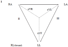
Figure 3: Einthoven triangle with correct lead placement.
The prevalence of ECG lead misplacement varies according to the acuity of the patient’s condition. Bond et al reported that the prevalence of lead misplacement in ECGs recorded in an intensive care unit (4%) was ten times higher than those recorded in an outpatient setting (0.4%) [6]. The2010 American Heart Association guideline for the management of Acute Coronary Syndrome (ACS) states that ECGs should be performed within ten minutes of arrival to hospital on all patients who present with possible ACS [7]. This target time, together with the severity of the patients’ condition, has the potential to result in even high errates of lead misplacement.
The ECG appearances with limb lead misplacement can easily and consistently be explained using the Einthoven triangle. The classical appearance of right arm- right leg misplacement is the presence of a near-flat line in lead II [8] while in right arm-left arm misplacement negative P-QRS-T complexes in leads I and a VL and positive QRS complexes in lead a VR are seen [9] (Figure 4 and 5).
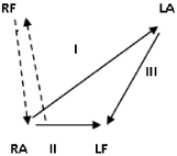
Figure 4: Right arm-left arm misplacement.
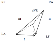
    Figure 5:  Right arm-left arm misplacement in an ECG with right arm-right leg
misplacement.
The most commonly encountered electrode misplacement is right arm-left arm misplacement, [4] which accounts for 20%of all cases [9,10]. ECG findings due to electrode misplacement can be difficult to detect despite producing characteristic appearances in many cases [11]. Despite the potential consequences of lead misplacement few ECG interpretation books cover adequately cover this topic [1,12].
In the process of reproducing these two misplacements we determined that the right arm - right leg was misplaced first followed by the right arm-left arm. When the leads were misplaced in the reverse order the near flat line in lead II moved to lead III. It despite a detailed literature search we were unable to identify any reports in which two limb lead misplacements occurred simultaneously.
Healthcare professionals responsible for recording and analyzing ECGs should receive adequate training to minimize the likelihood of incorrect lead placement. Thaler et al. achieved a 75% reduction in electrode misplacements in their intensive care unit through demonstration of errors made and a 45 min teaching session on electrode misplacement [13].
It is important to have easy access to previously recorded ECGs to facilitate timely comparison and recognition of errors. The ECG changes should be interpreted in light of the clinical history and all ECGs should be “signed off” by an adequately trained member of staff. Where lead misplacement is suspected, a new 12-lead ECG should be obtained and the one containing the error discarded or stored separately and used for training. Use of other resources such as the “REVERSE mnemonic” [14] should be considered as it has the potential to highlight incorrect lead placement (Table 1).
    Abnormal finding
    Significance


    R
    R wave is positive in a VR (P wave also is    positive)▲
    RA- LA lead misplacement


    E
    Extreme axis deviation: QRS axis is between +180    and -90 degrees
    RA- LA lead misplacement


    V
    Very low voltage (< 1mm) in isolated limb lead
    RL misplacement


    E
    Exchanged amplitude of P waves (P wave in lead I    is larger than in lead II) †
    LA- LL misplacement


    R
    R wave-abnormal progression in the precordial    leads●
    Precordial lead reversal


    S
    Suspect dextrocardia (negative P waves in lead I)
    Possible dextrocardia


    E
    Eliminate noise and interference (artefact    mimicking tachycardia or ST- changes)
    Artefact


      RA – right arm, LA – left arm, LL – left leg, RL    – right leg,
      ▲ Negative P-QRS-T in lead I,
      Concordant negative QRS and T waves in leads I,    II, III, a VL and/or a VF (RA – LL, RA – LA and RL – LL misplacement),
      † Sensitivity 90%15,
      ● The orientation of the QRS complexes in leads I    and V6 should be the same [8].
Table 1: “REVERSE” Mnemonic for common lead reversals and their significance [14].
Conclusion
ECG lead misplacement can alter the ECG appearance dramatically and represents an avoidable risk in patient management. A senior doctor should review all ECGs and together with education and training in recognition of lead misplacements this should reduce the prevalence of ECG lead misplacements.
References
- Lynch R. ECG interpretation: it’s easier when you know how. Global Emergency Care Skills Publishing. 2013.
- Eldridge J, Richley D, Baxter S, Blackman S, Breen C, Brown C, et al. Recording a standard 12-lead electrocardiogram. An approved methodology by the Society of Cardiological Science and Technology (SCST). Clinical guidelines by consensus. 2014.
- Rudiger A, Schöb L, Follath F. Influence of electrode misplacement on the electrocardiographic signs of inferior myocardial ischemia. Am J Emerg Med. 2003; 21: 574-577.
- Rudiger A, Hellermann JP, Mukherjee R, Follath F, Turina J. Electrocardiographic artifacts due to electrode misplacement and their frequency in different clinical settings. Am J Emerg Med. 2007; 25: 174-178.
- Jowett NI, Turner AM, Cole A, Jones PA. Modified electrode placement must be recorded when performing 12-lead electrocardiograms. Postgrad Med J. 2005; 81: 122-125.
- Bond RR, Finlay DD, Nugent CD, Breen C, Guldenring D, Daly MJ. The effects of electrode misplacement on clinicians' interpretation of the standard 12-lead electrocardiogram. Eur J Intern Med. 2012; 23: 610-615.
- O’Connor RE, Al Ali AS, Brady WJ, Brooks SC, Diercks D, Ghaemmaghami CA, et al. Part 9: Acute Coronary Syndromes: 2015 American Heart Association Guidelines for cardiopulmonary resuscitation and emergency cardiovascular care. Circulation. 2015; 132: s483-s500.
- Greenfield JC, Rembert JC. Mechanisms of very-low-voltage waveforms in either lead I, II, or III. J Electrocardiol. 2009; 42: 233-234.
- Harrigan RA, Chan TC, Brady WJ. Electrocardiographic electrode misplacement, misconnection, and artifact. J Emerg Med. 2012; 43: 1038-1044.
- Kligfield P, Gettes LS, Bailey JJ, Childers R, Deal BJ, Hancock EW, et al. Recommendations for the standardization and interpretation of the electrocardiogram. Part I: The electrocardiogram and its technology. A scientific statement from the American Heart Association Electrocardiography and Arrhythmias Committee, Council on Clinical Cardiology; the American College of Cardiology Foundation; and the Heart Rhythm Society. Endorsed by the International Society for Computerized Electrocardiography. J Am Coll Cardiol. 2007; 49: 1109-1127.
- Lynch R. ECG lead misplacement: A brief review of limb lead misplacement. Afric J Emerg Med. 2014; 4: 130-139.
- Crawford J, Doherty L. Practical Aspects of ECG Recording. M&K Update Ltd. 2012.
- Thaler T, Tempelmann V, Maggiorini M, Rudiger A. The frequency of electrocardiographic errors due to electrode cable switches: a before and after study. J Electrocardiol. 2010; 43: 676-681.
- Baranchuk A, Shaw C, Alanazi H, Campbell D, Bally K, Redfearn DP, et al. Electrocardiography pitfalls and artifacts: the 10 commandments. Crit Care Nurse. 2009; 29: 67-73.
- Abdollah H, Milliken JA. Recognition of electrocardiographic left arm/left leg lead reversal. Am J Cardiol. 1997; 80: 1247-1249.