Research Article
Austin Emerg Med. 2016; 2(7): 1039.
Investigation of Abnormal Findings in Lumbosacral MRI of Patients with Spondylolisthesis
Lotfinia I1, Daghighi MH2, Salehpour F1, Rezakhah A1, Mirzaei F1, Mahdkhah A1 and Naseri Alavi SA1*
1Department of Neurosurgery, Faculty of Medicine, Tabriz University of Medical Sciences, Tabriz, Iran
2Department of Radiology, Faculty of Medicine, Tabriz University of Medical Sciences, Tabriz, Iran
*Corresponding author: Seyed Ahmad Naseri Alavi, Department of Neurosurgery, Faculty of Medicine, Tabriz University of Medical Sciences, Tabriz, Iran
Received: August 06, 2016; Accepted: August 29, 2016; Published: August 31, 2016
Abstract
Introduction: Spondylolisthesis is a common disorder in lumabar vertebras and consist of more than 30% of lumbar fusions are diagnosed. Degenerative spondylolisthesis is seen more in women at level L4-L5 however isthmic spondylolisthesis is seen in men at level L5-S1. MRI is used commonly as first paraclinic test for evaluation of patients with back pain with or without radiculoapthy. It is used in supine position commonly that this cause glide at the reduced rate and along with a placement. This has to be misdiagnosed of Spondylolisthesis. The aim of this study is to investigate the MRIs of patients with Spondylolisthesis to explain the findings on MRI in these patients.
Methods and Materials: In this retrospective study all patients with spondylolisthesis that diagnosed by functional x-ray without report of radiologist note from January 2013 - January 2015 enrolled to the study.
Results: All the 85 patients have spondylolisthesis Grade 1 on MRI. Height of Disc, type of herniation, modic changes, existence of fluid or air in facet joint and existance of tropism in facet joint was investigated. Investigation of facet joint hypertrophy and facet tropism demonstrate no significant relation. Investigation of height of disc and disc intensity demonstrate no significant relation too.
Conclusion: Based on findings of this study we can realized that more patients with spondylolisthesis grade 1 on MRI misdiagnosed and this study help neurosurgeons to be suspected to spondylolisthesis with investigation of factors simultaneous such as disc protrusion, facet joint topism and flavum ligament hypertrophy.
Keywords: Spondylolisthesis; Modic change; Disk height; MRI
Abbreviations
CT: Computed Tomography; MRI: Magnetic Resonance Imaging; PET: Positron Emission Tomography
Introduction
Spondylolisthesis fused by a Belgium gynecologist about 200 years ago at first. Spondylolisthesis means glide to vertebrae on the lower one. This word is derived from two Greek word; Spondylos means vertebrae and listhesis means glide [1]. Spondylolisthesis is a common disorder in lumabar vertebras and consist of more than 30% of lumbar fusions are diagnosed. Spondylolisthesis is divided to 5 groups: Dysplastic, Isthmic, degenative, Traumatic and pathologic [2]. Degenerative spondylolisthesis is seen more in women at level L4- L5 however isthmic spondylolisthesis is seen in men at level L5-S1 [3]. In isthmic spondylolisthesis displacement is due to defects in the pars interarticularis; however in Degenerative spondylolisthesis there is no defect in pars interarticularis. The first symptom in these patients is back pain that exacerbate with exercise [4,5]. MRI is used commonly as first paraclinic test for evaluation of patients with back pain with or without radiculoapthy; however standard diagnosis is with lateral and felexion-extention graphy [6]. MRI can show soft tissue such as neural elements, disk herniaition, annulus defects, neoplastic or inflammation condition [7]. In most cases MRI is used for supine and this will allow it to glide vertebrae in the fall and along with being a vertebra diagnosis is not done correctly [7,8]. The aim of this study is to investigate MRI findings in patients with spondylolisthesis that diagnosed only with clinical findings.
Methods and Materials
After being approved by the ethics committee of Tabriz University of Medical Sciences, this descriptive study was performed in neurosurgery department in a 24 month period of time (January 2013-december 2015). Written informed consents were obtained from patients before enrollment. In this retrospective study all patients with Spondylolisthesis that diagnosed by functional x-ray but without report of it at radiologist note enrolled to the study. All patients referred to us for lumbar disk herniation or canal stenosis by progressive neurological symptoms. We investigate age, gender, disk intensity, type of herniation, modic change, existence of fluid or air in facet joint, facet tropism, hypertrophy of ligament flavum, level of spondylolisthesis, type and grade of spondylolisthesis and height of disk on MRI at admission. Exclusion criteria: patients with lumbar trauma, patients with past surgical history, patients with lumbar deformities.
Statistical analysis
Kruskal Wallis H and Mann Whiteny U (categorical data) were used for comparisons analysis. Statistical analysis was performed using SPSS software (USA). P value = 0.05 was regarded statistically significant.
Results
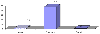
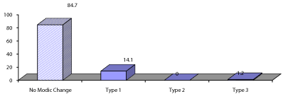
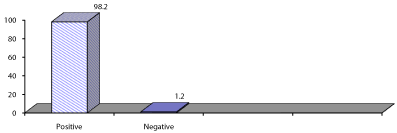
Eighty five patients with spondylolisthesis enrolled to the study. Eighty (94.1%) was female and 5 (5.9%) was male. The mean age was 48.29 ± 9.84 (Max = 75, Min = 28). Investigation of disk intensity is shown in Figure 1. Investigation of type of herniation showed that more patients had protrusion (Figure 2). Investigation of modic changes is shown in Figure 3. From 85 patients more patients had no air or fluid in facet joint (Figure 4). More than half of patients had facet tropism and 98.2% of patients had hypertrophy of ligament flavum (Figure 5,6). All the patients had spondylolisthesis grade 1, in more patients the level of involvement was L4-L5 and had degenerative spondylolisthesis (Figure 7,8). Investigation of disk height showed that more patients had normal height (Figure 9) and there was no relation between disk height and disk intensity (p-value = 0.40). There was no relation between facet tropism and hypertrophy of ligament flavum (p-value = 0.26).

    Figure 1:  Investigation of disk intensity on MRI. The number shows
frequencies (%).

    Figure 2:  Investigation of type of herniation on MRI. The number shows
frequencies (%).

    Figure 3:  Investigation of Modic change on MRI. The number shows
frequencies (%).

    Figure 4:  Investigation of existence of fluid or air in disk on MRI. The number
shows frequencies (%).

    Figure 5:  Investigation of facet tropism on MRI. The number shows
frequencies (%).

    Figure 6:  Investigation of hypertrophy of ligament flavum on MRI. The
number shows frequencies (%).

    Figure 7:  Investigation of level of disk involvement on MRI. The number
shows frequencies (%).

    Figure 8:  Investigation of type of spondylolisthesis on MRI. The number
shows frequencies (%).

    Figure 9:  Investigation of disk height on MRI. The number shows frequencies
(%).
Discussion
In this study, we demonstrate that there was no significant relation between disk intensity and disk height and also facet tropism and hypertrophy of ligament flavum. 85 patients with spondylolisthesis enrolled to study and investigated based on their MRI at admission. Some biomechanical studies proved that facet joint disorders and disk degenerative disease in lumbosacral joints cause degenerative spondylolisthesis [9-12]. Lumbosacral MRI is a routine choice for back pain complaints; however in supine position is not be usable. Axial MRI T2 is another choice help neurosurgeons diagnose existence of fluid or air in facet joints [13-16]. Mailleux et al demonstrated that existence of fluid on MRI T2 can help diagnosis of spondylolisthesis [17]. Caterini et al demonstrated that 66% of patients with spondylolisthesis had fluid in facet joints on MRI T2 however; 13.4% of patients without spondylolisthesis had fluid in facet joints too [18]. Investigation of fluid in facet joints in patients of this study was lower than them and was 31.8%. Based on results all the patients had grade 1 spondylolisthesis; most of them were female, most patients had protrusion herniation, most of them had hypertrophy of ligament flavum, most patients had L4-L5 involvement. These criteria can help neurosurgeons to be suspected to diagnosis of spondylolisthesis in early stages. In other hand half of patients had facet tropism that a good factor for diagnosis. In conclusion based on findings of this study we realized that more patients with spondylolisthesis grade 1 on MRI misdiagnosed and investigating of factors simultaneous such as disc protrusion, facet joint topism and flavum ligament hypertrophy help neurosurgeons to be suspected to spondylolisthesis.
References
- Kilian AJ, Picquet Ch. Dictionnaire geographique universal. Paris chez les editeures. 10: 1823-1833.
- Winn R.Yoman’s neurological surgery. 6th Ed, Saunders. 2011.
- Blackburne JS, Velikas EP. Spondylolisthesis in children and adolescents. J Bone Joint Surg Br. 1997; 59: 490-494.
- Frennered AK, Danielson BI, Nachemson AL. Natural history of symptomatic isthmic low-grade spondylolisthesis in children and adolescents: a seven-year follow-up study. J Pediatr Orthop. 1991; 11: 209-213.
- Gill GS, Manning JG, White HL. Surgical treatment of spondylolisthesis without spine fusion; escision of the loose lamina with decompression of the nerve roots. J Bone Joint Surg Am. 1955; 37: 493-520.
- Hresko MT, Labelle H, Roussouly P, et al. Classification of high-grade spondylolisthesis based on pelvic version and spine balance: possible rationale for reduction. Spine. 2007; 32: 2208-2213.
- Wiltse LL, Winter RB. Terminology and measurement of spondylolisthesis. J Bone Joint Surg Am. 1983; 65: 768-772.
- Wiltse LL, Newman PH, Macnab I. Classification of spondylolysis and spondylolisthesis. Clin Orthop Relat Res. 1976; 117: 23-29.
- Fujiwara A, Lim T, An HS, et al. The effect of disc degeneration and facet joint osteoarthritis on segmental flexibility of the lumbar spine. Spine. 2000; 23: 3036–3044.
- Schellinger D, Wener L, Ragsdale BD, Patronas NJ. Facet joint disorders and their role in the production of back pain and sciatica. Radiographics. 1987; 7: 923–944.
- Adams MA, Hutton WC. The mechanical function of the lumbar apophyseal joints. Spine. 1983; 8: 327–330.
- Yang KH, King AI. Mechanism of facet load transmission as a hypothesis for low back pain. Spine. 1984; 9: 557–565.
- Rihn JA, Lee JY, Khan M, et al. Does lumbar facet fluid detected on magnetic resonance imaging correlate with radiographic instability in patients with degenerative lumbar disease? Spine. 2007; 14: 1555–1560.
- Chaput C, Padon D, Rush J, et al. The significance of increased fluid signal on magnetic resonance imaging in lumbar facets in relationship to degenerative spondylolisthesis. Spine. 2007; 17: 1883–1887.
- Schinnerer KA, Katz LD, Grauer JN. MR findings of exaggerated fluid in facet joints predict instability. J Spinal Disord Tech. 2008; 21: 468–472.
- Cho BY, Murovic JA, Park J. Imaging correlation of the degree of degenerative L4–5 spondylolisthesis with the corresponding amount of facet fluid. J Neurosurg Spine. 2009; 11: 614–619.
- Mailleux P, Ghosez JP, Bosschaert P, et al. Distension ofthe inter-facet joints in MRI: an indirect sign of existing underestimationof spondylolisthesis and canal stenosis. J Belge Radiol. 1998; 81: 283–285.
- Caterini R, Mancini F, Bisicchia S, Maglione P, Farsetti P. The correlation between exaggerated fluid in lumbar facet joints and degenerative spondylolisthesis: prospective study of 52 patients. J Orthopaed Traumatol. 2011; 12: 87–91.