Case Presentation
Austin Emerg Med. 2016; 2(9): 1047.
Artery of Percheron Infarct with a Normal Initial MRI: A Case Report
Dias Ayesha M¹* and Rekha²
¹Department of Emergency Medicine, St John’s Medical College Hospital, Bangalore, India
²Department of Radiology, St John’s Medical College Hospital, Bangalore, India
*Corresponding author: Ayesha Maria Dias, Department of Emergency Medicine, St John’s Medical College Hospital, Bangalore, India
Received: November 13, 2016; Accepted: December 07, 2016; Published: December 09, 2016
Abstract
The artery of Percheron is a rare anatomical variant which supplies both the paramedian thalamic and the rostral midbrain. This unique feature is responsible for bilateral thalamic and mesencephalic infarctions if occluded. A single occlusion can lead to a clinical picture of drowsiness and visual disturbances due to the bilateral nature of the structures supplied.
We report a case of a patient in altered sensorium with normal early imaging (CT and MRI). Ischemia in the region of the artery of Percheron was revealed three days later with a repeat Magnetic Resonance Imaging. This is one of the few cases reported where early imaging fails to detect an acute stroke in a symptomatic patient.
Keywords: Artery of Percheron; Stroke; Thalamus; Coma
Introduction
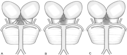
The thalamus receives a complex blood supply from the anterior, posterior, inferolateral and paramedian arteries [1,2]. The paramedian thalamic arteries can arise from the P1 segment of the posterior cerebral artery in three different ways. The commonest variation is when it arises from the P1 segment of each PCA. If the perforating arteries arise from a single common trunk which supplies both thalami across the midline this variation is the artery of Percheron (Figure 1). Occlusion of this artery produces paresis of upward gaze, drowsiness and often coma [3]. Imaging modalities are pivotal in identification for thrombolysis in acute stroke.

    Figure 1:  Variations of the paramedian thalamic-mesencephalic arterial
supply according to Percheron. A: In the most common variation, there are
many small perforating arteries arising from the P1 segments of the PCA. B:
The artery of Percheron is a single perforating blood vessel arising from one
P1 segment. C: The third type of variation is that of an arcade of perforating
branches arising from an artery bridging the P1 segments of both PCAs.
We describe a patient admitted with coma and normal early imaging by both CT and MRI which subsequently revealed a bilateral paramedian thalamic infarct.
Case Presentation
A forty four year old lady was brought to the emergency department with sudden loss of consciousness at her home half an hour after complaining of headache that evening. The family members accompanying her reported that she was drowsy for a short period before completely losing consciousness. She was initially managed at a local clinic where she was found to have a Glasgow coma scale (GCS) score of eight with localizing pain being her best motor response, uttering incomprehensible sounds and no visual response. Her pupils were noted to be 4mm bilateral sluggishly reactive. Her baseline investigations including a complete blood count, renal and liver parameters were all found to be within the normal range. She was in sinus rhythm and a formal echocardiography revealed a concentric left ventricular hypertrophy. The initial MRI and CT scan both done within twelve hours of presentation were both found to be normal. She was treated empirically with antiepileptic drugs and calcium channel blockers (Amlodipine), but as she was not responding to the treatment was referred to our emergency department (ED).
In our ED she had a blood pressure of 140/84 mmHg with a heart rate of 90/min. Her sensorium continued to be low (GCS - E2V3-4M4) on the second day of her illness and she was admitted to the intensive care unit. A doubtful history of fever five days prior to admission was obtained and a lumbar puncture done which showed no white blood cells and cultures were sterile. Further investigations including toxicology screen was also found to be negative.
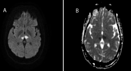
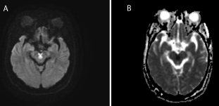
As her sensorium continued to fluctuate and her pupils remained sluggishly reactive and mid dilated during the next day she underwent a repeat MRI brain scan on day 3 of her illness. This revealed an acute infarct with diffusion restriction in bilateral anterior thalamic region (Figure 2) and interpeduncular regions (Figure 2) suggestive of an infarct in the artery of Percheron territory. She was started on anticoagulation and anti-platelets. Modafinil was also added to promote wakefulness in her.

    Figure 2:  Bilateral thalami infarct A. Diffusion restriction image B
Corresponding ADC image.

    Figure 3:  Interpeduncular infarct A. Diffusion restriction image B
Corresponding ADC image.
Her neurological condition improved from an impaired conscious state with non-reactive pupils bilaterally. She gradually was able to open her eyes and responded well to oral commands. Over the next few weeks in the neurology ward she improved and was discharged home in good condition with minimal memory deficits.
Discussion/Conclusion
Bithalamic paramedian infarcts are rare, and very difficult to suspect because of their complex anatomy, causing a large clinical variation [1]. The clinical presentation especially can be quite unusual with patients presenting with disorientation, memory impairment, and visual disturbances including vertical gaze palsy. The bilateral nature of the infarct produces these unique diffuse presenting features. A fluctuating consciousness level and impairment in arousal can be a characteristic finding especially within the first few days [3]. Very often an undifferentiated coma could be the only presenting feature developing within the first few days.
Lucia Rivera published a case report of a patient who presented with unusual behavior, minimal visual disturbances who suddenly became comatose needed intubation and ventilation only to regain consciousness after 2 days [4]. The MRI scan showed a bilateral thalamic infarct suggestive of an Artery of Percheron territory infarct.
A majority of cases reported, describe bilateral paramedian thalamic strokes clinically characterized by a triad of altered mental status, vertical gaze palsy, and memory impairment [5]. Altered mental status being described as a spectrum from drowsiness or confusion to hyper somnolence or coma.
Most of the cases reported have a presenting neurological image positive for a bilateral paramedian thalamic infarct. A diagnosis of an acute stroke and the benefit of thrombolysis makes it imperative to identify these cases early. MRI diffusion weighted images showing restriction in the area of the bilateral paramedian thalamic regions with normal T2 weighted images in the area of the artery of Percheron confirms an acute stroke. Our patient uniquely had an initial brain imaging which was normal on presentation. One similar case has been reported by Guillaume Cassourret et al. [6] a few reasons can account for this disparity. The lesion of occlusion being in the area of the paramedian thalamus, was deep and of small size. Both characteristics have been found to have a high risk of false negative reporting on early MRI scanning. In our case as the initial MRI scan was negative an angiography was not indicated. In conclusion our case clearly demonstrates that the role of MRI DWI in detecting an acute stroke though invaluable in ruling in an acute stroke may need to be repeated after 48hrs if all other aetiology for coma have been excluded and an artery of Percheron territory infarct is in fact a possibility even with a normal initial MRI scan.
From a more practical aspect, it is important to recognize that the clinical features though suggestive of an involvement of multiple vascular territories can be due to this unique variation. Furthermore it is wise to recognize this normal variation early when met with the possibility of bilateral median thalamic infarcts.
References
- Cassourret G, Prunet B, Sbardella F, Bordes J, Maurin O, Boret H. Ischemic stroke of the artery of Percheron with normal initial MRI: a case report. Case reports in medicine. 2010; 2010.
- Percheron G. The anatomy of the arterial supply of the human thalamus and its use for the interpretation of the thalamic vascular pathology. Zeitschriftfür Neurologie. 1973; 205: 1-3.
- Longo D, Fauci A, Kasper D, Hauser S. Harrison’s Principles of Internal Medicine 18th edition. McGraw-Hill Professional. 2011.
- Rivera-Lara L, Henninger N. Delayed sudden coma due to artery of Percheron infarction. Archives of neurology. 2011; 68: 386-387.
- Lazzaro NA, Wright B, Castillo M, Fischbein NJ, Glastonbury CM, Hildenbrand PG, et al. Artery of percheron infarction: imaging patterns and clinical spectrum. American Journal of Neuroradiology. 2010; 31: 1283-1289.
- Raphaeli G, Liberman A, Gomori JM, Steiner I. Acute bilateral paramedian thalamic infarcts after occlusion of the artery of Percheron. Neurology. 2006; 66: E7.
- Krampla W, Schmidbauer B, Hruby W. Ischaemic stroke of the artery of Percheron (2007: 10b). European radiology. 2008; 18: 192-194.
- Gentilini M, De Renzi EN, Crisi G. Bilateral paramedian thalamic artery infarcts: report of eight cases. Journal of Neurology, Neurosurgery & Psychiatry. 1987; 50: 900-909.
- Almamun M, Suman A, Arshad S, Kumar SJ. A Case of Midbrain and Thalamic Infarction Involving Artery of Percheron. Journal of Clinical Medicine. 2015; 4: 369-374.