
Special Issue - Intensive Care
Austin Emerg Med. 2021; 7(1): 1070.
Tension Pneumothorax due to Colon Perforation in Delay Post Traumatic Diaphragmatic Hernia: Case Report
Samsami M, Tahmasbi H, Nikraftar P and Bagherpour JZ*
Department of Surgery, Shahid Beheshti University of Medical Sciences, Imam Hossein Hospital, Iran
*Corresponding author: Javad Zebarjadi Bagherpour, Department of General Surgery, Shahid Beheshti University of Medical Sciences, Imam Hossein Hospital, Shahid Madani Avenue, Imam Hossein Square, Tehran, Iran
Received: March 30, 2021; Accepted: April 22, 2021; Published: April 29, 2021
Abstract
Diaphragmatic injuries were described first by Sennertus in 1541. Rupture of the diaphragm due to blunt trauma is a rare event that is usually not detected in the acute phase of trauma and may manifest itself late and with dangerous complications. The common side effects of this injury include displacement of the abdominal viscera into the thoracic cavity, which can cause respiratory problems due to limited lungs. Abdominal organs such as stomach, omentum, intestines, spleen, and liver are the most common to herniate in to the thoracic cavity .In late presentation, the key point is to identify the patient’s strong clinical suspicion and history. CT scan is the most common modality in diagnosis of diaphragmatic hernia. In this article, we introduce a unique case of diaphragmatic hernia after trauma due to falling from a height of 2 years ago, which showed itself with a tension pneumothorax in its management.
Keywords: Diaphragmatic hernia; Complication; Trauma
Introduction
Diaphragmatic injury may occur due to both penetrating and blunt traumas [1]. The frequency with which either of these mechanisms leads to diaphragmatic injury varies according to the geographic location [2]. The incidence of post-traumatic blunt diaphragmatic injury varies from 0.16 to 5%. Up to 70% of diaphragmatic tears are missed initially [3]. The most common causes of diaphragmatic hernia due to blunt injuries include falling from a height and road accidents; rate of diaphragmatic hernia mortality varies from 5 to 51% [4].The common side effects of this injury include displacement of the abdominal viscera into the thoracic cavity, which can cause respiratory problems due to limited lungs. Other dangerous side effects include strangulation of viscera inside the thorax, which can be up to 70% mortality rate. Diaphragmatic hern diagnosis methods include chest x-ray, MRI, and CT scan, which is the most common modality [5]. Approximately 69% of traumatic hernias are left sided, 24% are right- sided and 15% are bilateral. Diaphragmatic hernia diagnosis miss in 56% of cases in the acute phase if it is asymptomatic [6]. High index of clinical suspicion is required because of the missed diagnosis and potential for delayed presentation. Here we introduce a case of diaphragmatic hernia after blunt trauma, which was referred with tension pneumothorax due to necrosis of the colon inside the thorax cavity.
Case Presentation
A 52-year-old male was admitted to the emergency department with fever and dyspnea from 2 days ago. Initially the vital sign was pulse rate = 110, respiratory rate = 32, oxygen saturation = 83%, Temperature = 38 and blood pressure = 130/70. In the patient’s history, there was a history of femoral fractures due to a fall from a height of 6 meters 2 years ago. At the CT-scan there was a pneumothorax in the left hemithorax with shifting mediastinum. And Findings in favor of diaphragmatic hernia (Figure 1A-1C). Chest tube was inserted in left hemithorax. After insertion, gas with fecal material was drained from chest tube. The patient transferred to operation room after resuscitation with diagnosis colonic perforation due to diaphragmatic hernia. At first midline laparotomy was done. Transverse colon was herniated to thorax from a defect about 3 centimeters in left side diaphragm. Colon extracted from diaphragm. There was a perforation in transverse colon about 5 centimeters from splenic flexure. Colon released from spleen and after refreshing the perforation margin a loop colestomy was done from perforation site. Then, posterolateral thoracotomy was done. There was fecaloid material in pleural space and peel on lung. Washing and decortication was done, two chest tubes were inserted, and patient transferred to surgical intensive care unit, and antibiotic therapy was performed with broad-spectrum antibiotics. To prevent abscess formation and re-infection, Thorax cavity washes through continuous thoracic tubes. After 4 days, the patient was transferred to the ward, and after ensuring that the lungs were completely expand, the chest tubes were removed (Figure 1C). On the 10th day after the operation, he was discharged from the hospital. Two months later, he underwent colostomy clouser and ao complications were found in 6 month follow up.
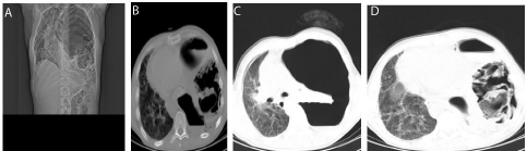
Figure 1A-1D: CT scan shows colon in hemithorax with pneumothorax.
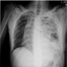
Figure 2: Post chest tube chest x-ray shows finding of diaphragmatic hernia.
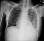
Figure 3: Post chest tube removal chest x-ray shows finding of diaphragmatic hernia.
Discussion
Diaphragmatic injuries were described first by Sennertus in 1541. Riolfi performed the first successful repair in 1886. Not until 1951, when Carter et al., published the first case series, was this injury well understood and delineated [7]. Abdominal organs such as stomach, omentum, intestines, spleen, and liver are the most common to herniate in to the thoracic cavity [8]. In our patient, part of the transverse colon was inside the thorax. Patients may therefore experience chest pain, recurrent shortness of breath, and gastrointestinal symptoms such as nausea and vomiting, epigastric discomfort, or abdominal pains and bowel sounds may be heard in left side of the chest [9]. In our patient, there was a history of active dyspnea as well as changes in bowel habits as occasional diarrhea and intermittent abdominal pain. As mentioned earlier in the introduction, the most important diagnostic modality for Herni is CT scan aperture. Common findings on CT scans include direct visualization of injury, Segmental diaphragm non-visualization, Intrathoracic herniation of viscera, peridiaphragmatic active contrast extravasation [10]. Treatment of diaphragmatic hernia is surgical via laparotomy or thoracotomy. Acute cases are better managed via a laparotomy as this also rules out and treats associated intraabdominal organ injuries. Delayed cases, however, are better treated via a thoracotomy or thoracoabdominal approach because of intrathoracic adhesions [11]. In our patient, laparotomy was performed first however no adhesion was encountered between the herniated organs and the lungs despite presenting after 2 years of the blunt abdominal injury, and after finding the pathology, the transvers loop colostomy was implanted for the patient from the transverse colon perforation site, and then thoracotomy was performed. The thoracic cavity was flushed, the diaphragm was repaired, and two chest tubes were inserted. Due to the contamination of the thorax cavity with feces, our concern was the formation of abscesses and reinfection, which was continuously the washing of the thorax cavity through the thoracic tubes. Fortunately, no side effects were reported.
Conclusion
In delayed diaphragmatic hernias, a history of previous trauma as well as a strong clinical suspicion are necessary to diagnose and use radiological instruments to confirm the diagnosis, and surgical treatment should be performed as soon as the diagnosis is made to prevent complications.
References
- Kidmas A, et al. Delayed presentation of blunt traumatic diaphragmatic hernia: a case report. Nigerian journal of surgical research. 2005; 7: 323-324.
- Adler DH. Blunt diaphragmatic injury in a 7-year-old girl. The Journal of emergency medicine. 2002; 23: 39-42.
- Davoodabadi A, et al. Blunt traumatic hernia of diaphragm with late presentation. Archives of trauma research. 2012; 1: 89-92.
- Edino S, S Alhassan and O Ajayi. Traumatic diaphragmatic rupture with gastro-pleuro-cutaneous fistula: A case report and literature review. Nigerian J Surg. 2002; 8: 18-20.
- El-Yakub A, et al. Delayed presentation of posttraumatic diaphragmatic hernia masquerading as recurrent acute asthmatic attack. Case reports in medicine, 2017. 2017.
- Ganie FA, et al. The characteristics and surgical approach in post-traumatic diaphragmatic hernia: a single center experience. Bulletin of Emergency & Trauma. 2013; 1: 108-111.
- Sachdeva R, S Sachdeva and S Solanki. Acquired diaphragmatic hernia in an adult male: A diagnostic challenge. Nepal Journal of Medical Sciences. 2013; 2: 194-196.
- Mansoor E. Post-traumatic diaphragmatic hernias-importance of basic radiographic investigations. South African Journal of Surgery. 2013; 51: 75- 76.
- Ozdemir S, et al. Late complication of diaphragmatic injury: hernia-a report of 4 cases and review of literature. Annals of Gastroenterology, 2009; 22: 275-277.
- Sliker CW. Imaging of diaphragm injuries. Radiologic Clinics. 2006; 44: 199- 211.
- Kishore G, et al. Traumatic diaphragmatic hernia: tertiary centre experience. Hernia. 2010; 14: 159-164.