Research Article
Austin J Endocrinol Diabetes. 2014;1(6): 1027.
Sonographic Features Suggestive of Papillary Thyroid Carcinoma
Sheikh HI1, Toubi AU2, Shechner CA3 and Saiegh LE1,3*
1Department of Internal Medicine B, Bnai-Zion Medical Center, Israel
2Ultrasound unit, Radiology Department, Bnai-Zion Medical Center, Israel
3Endocrinology Departments, Bnai-Zion Medical Center, Israel
*Corresponding author: Saiegh LE, Endocrinology Department, Bnai-Zion Medical Center, Israel,
Received: August 08, 2014; Accepted: October 09, 2014; Published: October 10, 2014
Abstract
Aim: To examine the prevalence and the diagnostic value of different ultrasonographic features in thyroid papillary carcinoma.
Methods: The sample of the study consisted of 74 patients, 40 of them have had the diagnosis of papillary cancer on ultrasound guided fine-needle-aspiration, while the other 34 patients have had the diagnosis of benign colloid nodule. The ultrasonographic images were re-reviewed and several features were evaluated and correlated with the final cytology.
Results: The ultrasonographic features that have been associated with malignancy were: non-homogenous hypo-echogenicity (P<0.017), poorly defined margins (P<0.008), presence of micro-calcifications (P<0.001) and lobular contour (P<0.05). The presence of colloid was associated with benignity (P<0.001). On the other hand, size of nodule, number of nodules and presence of macro-calcifications were not associated with papillary carcinoma.
Conclusions: In a thyroid nodule, the ultrasonographic features that were significantly associated with papillary cancer were: non-homogenous hypo-echogenicity, poorly defined margins, presence of micro-calcifications and lobular contour.
Keywords: Ultrasound; Fine-needle aspiration; Papillary cancer
Introduction
Thyroid nodule is a common finding. It could be revealed in approximately 4-8% of adult people by palpation and in 50% by post mortem histological examination. The prevalence of thyroid nodules increases with age [1]. Risk factors that predispose for thyroid cancer are: male gender, age below 20 or above 60, family history and childhood neck irradiation.
Papillary thyroid carcinoma (PTC) is the most common type of thyroid malignancy in children and adults, as it accounts for 80% of all thyroid malignancy cases.
Fine needle aspiration (FNA) is the most important initial diagnostic test of thyroid nodule. The accuracy of this test is between 70-97% [1]. Approximately 70% of tests are benign, 4% malignant, 10% suspicious and 10% of the tests are interpreted as non-conclusive [2].
Ultrasound (US) is an important tool for evaluation of a thyroid nodule. The current recommendation is to perform FNA with ultrasound guidance for nodules that can’t be palpated. But, many researches have shown that ultrasound guidance is important in palpable nodules as well, as it increases the accuracy of the test compared to FNA alone.
Many studies have been carried out in order to distinguish between benign and malignant nodules according to different ultrasonographic features, but characterization of nodules as either benign or malignant remains problematic because of considerable overlap in ultrasonographic features. Presence of micro-calcifications, hypo-echogenicity, nodule size, irregular borders, absence of “halo sign”, central vascularity, one nodule versus multiple and solid nodule versus cystic one, all have shown in some studies to be correlated with malignancy [3,4]. However, the sensitivity and specificity of these findings were not high enough. So, radiologic diagnosis and distinction between benign and malignant nodules are often not possible. These features are used mainly to guide the cytopathologist which nodule to examine by FNA in the cases when multiple nodules exist.
Although there have been multiple studies describing the ultrasonographic features of papillary thyroid carcinoma, very few have examined all the features in a single study.
The purpose of the current study is to examine the prevalence and the diagnostic value of different ultrasonographic features in thyroid papillary carcinoma.
Material and Methods
We retrospectively reviewed the ultrasonographic features of cases of papillary thyroid carcinoma and benign nodules imaged over a year (January 2012 to December 2012) at our institution (Bnai-Zion Medical Center, Haifa, Israel). Our study comprises 74 patients, 40 of them were diagnosed to have papillary cancer, while the other 34 had harbored a benign colloid nodule by cytology. Thyroid US was performed by a single skilled radiologist with GE Logiq 9 ultrasound machine using 10 MHz linear transducer. In real time, the radiologist recognized the nodule and the cytologist inserted the needle when the same radiologist still imaging the nodule. FNA was performed using a 23-guage needle, and in each case, an average of two passes was carried out for each nodule.
Re-reviewing the ultrasonographic images was performed in a “blind” manner, without any prior knowledge of the cytological interpretation of each case.
Different ultrasonographic features were evaluated in each image: The feature “echogenicity” was sorted as hypo echoic, hyper echoic, isoechoic, non homogeneous hypo echoic, non homogenous hyper echoic or non homogenous isoechoic nodule. The feature “echo texture” was sorted as dominant solid, dominant cystic or mixed nodule. We also evaluated the presence of hyper-echoic echogenecities such as micro-calcifications (defined as punctuate echogenic foci with or without acoustic shadowing). Macro-calcifications (defined as course irregular hype rechoic foci casting acoustic shadowing), colloid matter (tiny echo genic foci with a posterior comet-tail or ring down artifact). The feature “nodule margins” was sorted as well defined (with or without halo- defined as a thin rim of decreased echogenicity surrounding the neoplasm) or not defined. The feature “nodule size” was sorted as either less than 1cm, between 1-2cm, between 2-3cm, and more than 3cm. The feature “number of nodules” was sorted as one nodule versus multiple nodules. The feature “nodule contour” was sorted as symmetrical round, lobular or elongated in transitional section.
The prevalence of each feature was examined and was correlated with cytological interpretation.
Statistical analysis was carried out using the statistical software SPSS 15. Statistical comparison of the various parameters and features was performed by using the test 2χ and the student’s t-test. P-values less than 0.05 were considered statistically significant.
The study was performed according to the Declaration of Helsinki and approved by the institutional ethics committee.
Results
The sample of the study consisted of 74 patients, 40 (54.1%) of them have had the diagnosis of papillary cancer on US guided FNA, while the other 34 (45.9%) patients have the diagnosis of benign colloid nodule.
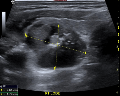
Regarding the ultrasonographic features: number of nodules, nodule size and macro-calcifications, the current study showed none to be significantly associated with papillary cancer. On the other hand, we found that the feature “non homogeneous hypo echoic” nodule (Figure 1) was significantly associated with malignancy with sensitivity of 92.5%, negative predictive value of 76.92% and a positive predictive value of 60.65% (P<0.017). The feature “poorly defined margins” (Figure 2) was also significantly associated with malignancy with sensitivity of 92.5%, specificity of 32.35%, negative predictive value of 78.57% and positive predictive value of 61.66% (P<0.008). A statistically significant association with malignancy was also observed for the presence of micro-calcifications (Figure 3) and lobular contour (Figure 2) with a specificity of 100% and 91.17% respectively, positive predictive value of 100% and 78.57% respectively (P<0.001, P<0.05 respectively).
Figure 1: 49 year old man with papillary thyroid carcinoma. Ultrasound revealed a non-homogenous hypo-echoic nodule compared to the echogenicity of the surrounding thyroid parenchyma.
Figure 2: Ultrasonographic image of a 56-year-old woman with a nonhomogenous hypo-echoic solid papillary thyroid carcinoma (white arrow) with poorly-defined margins and irregular, lobulated contours.
Figure 3: Punctuate echogenicities in a thyroid nodule. Micro-calcifications (white arrows-upper image): pinpoint punctuate areas of increased echogenicity measuring less than 1 mm with or without posterior acoustic shadowing. Macro-calcifications (white head arrow-upper image): areas of increased echogenicity measuring larger than 1 mm with posterior acoustic shadowing. It should be noticed that micro-calcifications and macrocalcifications should be distinguished from the colloid material that can give a stripped bright echogenic spotting with no posterior acoustic shadowing (white arrow-lower image).
The presence of colloid was associated significantly with benignity with specificity of 100% , sensitivity of 41.17%, positive predictive value of 100% and negative predictive value of 66.66% (P<0.001).
Discussion
Ultrasound is a highly sensitive tool to evaluate thyroid nodules. In cases with multiple nodules, it is important to determine which nodules to aspirate according to several suspicious Sonographic features. Some studies revealed that nearly half (48%) of PTC cases were present in the setting of a multi-nodular thyroid [5]. In our study, number of nodules was not significantly correlated with cytology. Thus, one shouldn’t assume that any given nodule is benign simply because it resides in a multi-nodular thyroid.
Concerning the feature “size of nodule “, several studies had found that there is no correlation between nodule’s size and malignancy. Benign nodules can reach a large size while PTCs can be too small [5]. Similar results were observed in the current study with no significant correlation between nodule’s size and papillary cancer. Therefore, biopsy of only the largest nodule is ill-advised.
The feature “echogenicity” is related to the echogenicity of a nodule compared to the echogenicity of the surrounding thyroid parenchyma. Some studies showed that 63-90% of papillary cancer is found in hypo echoic nodules [1, 6-9]. On the other hand, this sonographic feature is not specific enough because 55% of benign nodules were also hypo echoic. In our study we found that non-homogenous hypo echoic nodules are significantly associated with papillary cancer with high sensitivity of 92.5% while a low specificity of 29.4% (P<0.0017) which is consistent with previously reported data. Therefore, a non-homogenous hypo echoic nodule could be a suggestive finding of papillary cancer.
While some studies have found that most of papillary cancer nodules were solid nodules, and only 13% of papillary cancer nodules had a cystic component [10], in our study we had only few cases of cystic or mixed nodules, so we cannot conclude about the correlation of the feature “echo-texture” and malignancy.
Many studies have shown that existence of intra-nodular micro-calcifications is the most specific finding for malignancy, with specificity of 95%, while sensitivity of 29-59% [1, 6-9, 11, 12]. This is consistent with our findings which showed specificity of 100% and sensitivity of 55% (P<0.001). So, the presence of intra-nodular micro-calcifications is highly suggestive of papillary cancer. On the other hand, macro-calcifications did not correlate with malignancy in previous studies neither in the current study [13, 14].
The margins refer to the border separating a thyroid nodule from the surrounding parenchyma and on ultrasound can be either well defined or poorly defined depending on the extent of tumor invasion. In one series, the feature “poorly defined margins” was found in 77% of malignant nodules versus 15% of benign [6]. On the other hand, other studies showed that 50% of papillary cancer nodules are well defined nodules [1, 12, 15, 16]. In our study we found that poorly defined margins is significantly correlated with malignancy with sensitivity of 92.5% and specificity of 32.35% (P<0.008). The feature “lobular contour” was also significantly correlated with malignancy. This finding is most probably attributed to the fact that malignant tissue tends to grow unevenly and give the lobular contour [11]. We did not have enough cases with elongated nodules to obtain any conclusion about this feature.
Conclusions
The sample of the study consisted of 74 patients, 40 (54.1%) of them have had the diagnosis of papillary cancer on US guided FNA, while the other 34 (45.9%) patients have had the diagnosis of benign colloid nodule. Several ultrasonographic features were associated with malignancy, so they can be used to predict the risk of papillary cancer. The risk of malignancy is expected to be low in the absences of these features and conversely higher in their presence. None of these features can exclude malignancy, but clinicians can use them to estimate the risk of malignancy.
Features more suggestive of papillary cancer were a non-homogenous hypo echoic nodule with intra-nodular micro-calcifications, lobular contour and poorly defined margins. Those features can be used in patients with multi-nodular thyroid gland in order to decide which nodule to aspirate by FNA.
References
- Chan BK, Desser TS, McDougall IR, Weigel RJ, Jeffrey RB Jr . Common and uncommon sonographic features of papillary thyroid carcinoma. J Ultrasound Med. 2003; 22: 1083-1090.
- Cibas ES, Ali SZ; NCI Thyroid FNA State of the Science Conference . The Bethesda System For Reporting Thyroid Cytopathology. Am J Clin Pathol. 2009; 132: 658-665.
- Singer PA, Cooper DS, Daniels GH, Ladenson PW, Greenspan FS, Levy EG, et al . Treatment guidelines for patients with thyroid nodules and well-differentiated thyroid cancer. American Thyroid Association. Arch Intern Med. 1996; 156: 2165-2172.
- Tollin SR, Mery GM, Jelveh N, Fallon EF, Mikhail M, Blumenfeld W, et al . The use of fine-needle aspiration biopsy under ultrasound guidance to assess the risk of malignancy in patients with a multinodular goiter. Thyroid. 2000; 10: 235-241.
- Jun P, Chow LC, Jeffrey RB . The sonographic features of papillary thyroid carcinomas: pictorial essay. Ultrasound Q. 2005; 21: 39-45.
- Papini E, Guglielmi R, Bianchini A, Crescenzi A, Taccogna S, Nardi F, et al . Risk of malignancy in nonpalpable thyroid nodules: predictive value of ultrasound and color-Doppler features. J Clin Endocrinol Metab. 2002; 87: 1941-1946.
- Solbiati L, Volterrani L, Rizzatto G, Bazzocchi M, Busilacci P, Candiani F, et al . The thyroid gland with low uptake lesions: evaluation by ultrasound. Radiology. 1985; 155: 187-191.
- Watters DA, Ahuja AT, Evans RM, Chick W, King WW, Metreweli C, et al . Role of ultrasound in the management of thyroid nodules. Am J Surg. 1992; 164: 654-657.
- Rago T, Vitti P, Chiovato L, Mazzeo S, De Liperi A, Miccoli P, et al . Role of conventional ultrasonography and color flow-doppler sonography in predicting malignancy in 'cold' thyroid nodules. Eur J Endocrinol. 1998; 138: 41-46.
- Frates MC, Benson CB, Doubilet PM, Cibas ES, Marqusee E . Can color Doppler sonography aid in the prediction of malignancy of thyroid nodules? J Ultrasound Med. 2003; 22: 127-131.
- Kim EK1, Park CS, Chung WY, Oh KK, Kim DI, Lee JT, Yoo HS . New sonographic criteria for recommending fine-needle aspiration biopsy of nonpalpable solid nodules of the thyroid. AJR Am J Roentgenol. 2002; 178: 687-691.
- Koike E, Noguchi S, Yamashita H, Murakami T, Ohshima A, Kawamoto H, et al. Ultrasonographic characteristics of thyroid nodules: prediction of malignancy. Arch Surg. 2001; 136: 334-337.
- Khoo ML, Asa SL, Witterick IJ, Freeman JL . Thyroid calcification and its association with thyroid carcinoma. Head Neck. 2002; 24: 651-655.
- Kakkos SK, Scopa CD, Chalmoukis AK, Karachalios DA, Spiliotis JD, Harkoftakis JG, et al . Relative risk of cancer in sonographically detected thyroid nodules with calcifications. J Clin Ultrasound. 2000; 28: 347-352.
- Leenhardt L, Hejblum G, Franc B, Fediaevsky LD, Delbot T, Le Guillouzic D, et al . Indications and limits of ultrasound-guided cytology in the management of nonpalpable thyroid nodules. J Clin Endocrinol Metab. 1999; 84: 24-28.
- Propper RA, Skolnick ML, Weinstein BJ, Dekker A . The nonspecificity of the thyroid halo sign. J Clin Ultrasound. 1980; 8: 129-132.


