Special Article - Diabetes Mellitus
Austin J Endocrinol Diabetes. 2019; 6(1): 1065.
The Proportion of HCC Related to Obesity, Diabetes, and Metabolic Syndrome will Likely Increase in the Future; Non Alcoholic Steatohepatitis HCC
Hamed AK*
Department of Internal Medicine, Hepatology and Diabetes, Egyptian Military Medical Academy, Egypt
*Corresponding author: Abd Elkhalek Hamed, Department of Internal Medicine, Hepatology and Diabetes, Egyptian Military Medical Academy, Egypt
Received: March 26, 2019; Accepted: May 08, 2019; Published: May 15, 2019
Abstract
NAFLD is a wide spectrum disease ranging from simple steatosis, NASH to liver cirrhosis and is the most common type of CLD in the Western world (25% in the general population). Many studies showed an increased incidence of T2 DM in patients with NAFLD independently of ordinary risk factors. This association carries more burdens to CVD, also cancer including HCC, which happens particularly in those with NASH and exacerbated by metabolic syndrome. The pathophysiology of NAFLD involves many factors, and the intestinal microbiota plays an important role in the pathogenesis of both NAFLD and HCC. Although the risk ratios of DM, obesity, and metabolic syndrome do not approach those of HCV or HBV, they are far more prevalent conditions than HCV and HBV in developed countries. Given the increasing prevalence of these conditions, the proportion of HCC related to obesity, DM, and metabolic syndrome will likely increase in the future. Lack of a general consensus due to the paucity of data makes us still do not know if it is Possible to Stop or Cease the NASH to Turn into HCC and increase its incidence. However Exercise, specially vigorous, Metformin and Statins may be helpful. Obeticholic acid, Elafibranor, Selonsertib and Cenicriviroc, the new drugs for NAFLD/NASH treatment we are waiting for, may carry more hope to decrease HCC.
Keywords: Obesity; Diabetes; Metabolic Syndrome
Abbreviations
CLD: Chronic Liver Disease; NAFLD: Nonalcoholic Fatty Liver Disease; MS: Metabolic Syndrome; T2DM: Type2 Diabetes Mellitus; NASH: Non Alcoholic Steatohepatitis; HCC: Hepatocellular Cancer; TLR: Toll-Like Receptor, DAMP: Damage-Associated Molecular Patern; LPS: Lipopolysaccharide; PAMP: Pathogen-Associated Molecular Patern
Introduction
NAFLD is a wide spectrum disease ranging from simple steatosis, NASH to liver cirrhosis and is the most common type of CLD in the Western world. Many studies showed increased incidence of T2 DM in patients with NAFLD independently of ordinary risk factors. The risk of T2 DM ranged from 33% to 55% in patients with NAFLD [1].
A meta-analysis of 20 observational studies, involving more than 115 000 individuals, demonstrated that NAFLD was associated with an almost two-fold increased risk of T2 DM over a median period of 5 years [2].
The prevalence of NAFLD is estimated to be about 25% in the general population, and even higher in certain parts of the world and in some patient populations, such as among the obese and patients with diabetes. This high prevalence is closely associated with the rise in the rates of obesity and MS. Components of MS include increased fasting plasma glucose or T2DM, hypertriglyceridemia, low high density lipoprotein level, increased waist circumference, and hypertension [3].
Bidirectional relation between NAFLD and MS component was documented recently, most important is T2DM. This association carries more burdens to CVD, also cancer including HCC [1,4].
observational studies has shown that older age, too much fibrosis, diabetes, obesity, MetS, high iron, much alcohol consumption, and menopause are further risk factors for HCC development in NAFLD/ NASH [3].
HCC is rare among adolescents and accounts for less than 1% of all malignant neoplasms among children younger than 20 years [5].
The progression to HCC in NAFLD happens particularly in those with NASH and exacerbated by metabolic syndrome (Table 1), or PNPLA3 gene polymorphism [6].
    Study Type
    HCC Cases
    Metabolic Syndrome Definition
    RR (95% CI)


    Case-control
    3,649 cases of 195,953 controls
    NCEP-ATP III
    2.58 (2.40-2.76)


    Cohort
    266 cases of 578,700
    WHO
    1.35 (1.12-1.63)


    Cohort
    1,931 cases of 23,625
    NCEP-ATP III
    M:1.89 (1.11-3.22); F:3.67 (1.78-7.57)


    Cohort
    1,858 cases of 27,724
    AHA
    M:1.73 (1.03-2.91);F:1.18 (0.55-2.51)


    RR:    Risk Ratio
Table 1: Association Between Metabolic Syndrome and HCC: Systemic Review and Meta-Analysis-modified from [7].
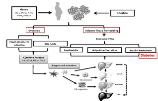
The pathophysiology of NAFLD involves many factors, and the intestinal microbiota plays an important role in the pathogenesis of both NAFLD and HCC. NAFLD and NASH are associated with alterations in the gut microbial population through various routes, such as alterations in gut epithelial permeability, choline metabolism, endogenous alcohol production, release of inflammatory cytokines, regulation of hepatic TLR, and bile acid metabolism. In addition, altered bile acid metabolism, release of inflammatory cytokines, and TLR-4 expression may promote NAFLD/NASH-associated HCC [8,9] (Figure 1).

Figure 1: Role of Obesity and MS in HCC Pathogenesis.
Obesity and metabolic syndrome lead the oncogenesis in the setting of abnormal hepatic morphology, and hepatic steatosis may provide the appropriate microenvironment for the development of cancer. Insulin resistance leads to fat accumulation in the hepatocytes by lipolysis and hyperinsulinemia. Obesity may lead to release of proinflammatory cytokines, inhibition of anti -inflammatory cytokines, and lipotoxicity.
Although the RRs of DM, obesity, and metabolic syndrome do not approach those of HCV or HBV (Table 2), they are far more prevalent conditions than HCV and HBV in developed countries. Given the increasing prevalence of these conditions, the proportion of HCC related to obesity, DM, and metabolic syndrome will likely increase in the future [10].
    Study Type
    HCC Cases
    Metabolic Syndrome Definition
    RR (95% CI)


    Case-control
    3,649 cases of 195,953 controls
    NCEP-ATP III
    2.58 (2.40-2.76)


    Cohort
    266 cases of 578,700
    WHO
    1.35 (1.12-1.63)


    Cohort
    1,931 cases of 23,625
    NCEP-ATP III
    M:1.89 (1.11-3.22); F:3.67 (1.78-7.57)


    Cohort
    1,858 cases of 27,724
    AHA
    M:1.73 (1.03-2.91);F:1.18 (0.55-2.51)


    RR:    Risk Ratio
Table 2: Multivariate analysis of variables associated with HCC. Modified from
[11].
HCC in NAFLD Non-cirrhosis
Has usually large measurements, is partially or well differentiated, and generally lacks encapsulation. HCC in NAFLD may arise in the absence of histologically definite or evident inflammation. Dysregulated hepatic and circulating pro-inflammatory cytokines and adipokines, oxidative and endoplasmic reticulum stress, and dysbiosis are likely associated with obesity-related Hepatocarcinogenesis [12].
An interesting possibility is the malignant transformation of hepatocellular adenoma, associated with obesity in non-cirrhotic patients with NAFLD [3].
Currently, there is a lack of recommendations for surveillance of patients with NASH and without cirrhosis, probably because of the difficulty of identifying patients with NASH without a liver biopsy. However, recent blood-based biomarkers could be an affordable alternative for identification of patients at high risk of NASH and advanced fibrosis [13].
The incidence and prevalence of HCC in NAFLD depend on the stage of underlying fatty liver disease. While the epidemiological data in relation to HCC in viral hepatitis and alcoholic hepatitis are consistent, there is a lack of strong epidemiological data concerning the incidence and prevalence of HCC in NAFLD. A few longitudinal outcome studies explored the prevalence of HCC in NASH, reporting a prevalence varying from 0 to 3% on a follow-up period between 5.6 and 21 years. The percentage was increased if the incidence of HCC in NAFLD cirrhosis was considered, with a cumulative HCC incidence ranging between 2.4% with a median follow-up of 7.2 years and 12.8% with a 3.2-year median follow-up [14].
Is that possible to stop or cease the NASH to turn into HCC and increase its incidance?
We do not exactly know this because the etiopathogenesis of NASH is not completely known. Primary prevention, which is our aim focuses on risk factors for HCC and their treatment, secondary prevention concentrates on the treatment of underlying liver diseases in patients with HCC aiming at a prevention of disease progression, and tertiary prevention aims at a reduction of recurrence after successful curative treatment of HCC [15].
A few chemopreventive agents have shown promise in the prevention and treatment of steatohepatitis and fibrosis (small individual studies ); thus there is a lack of a general consensus due to paucity of data. Exceptions include nucleoside analogues used to reduce hepatitis B viral replication, and DAAs for HCV which have very high cure rates [16].
Preventions and treatment in NAFLD-Related HCC
Lifestyle changes: Considering the pathogenesis of HCC in NAFLD, one has to face the burden of obesity and diabetes, lifestyle changes is the cornerstone for primary prevention and has been shown to reduce incident NAFLD and the related metabolic disorders and also may reverse NASH and liver fibrosis. It has been reported to have a preventive effect on the development of HCC. A protective effect in the development of HCC has been attributed in general to the Mediterranean diet.
Bariatric surgery could be recommended for patients with morbid obesity, it may reduce Liver fibrosis but carries a risk of decompensation in advanced cirrhotics. Observational studies and animal work suggest that exercise can prevent HCC development (inhibition of mTOR and the activation of AMPK, which are both involved in cell growth and proliferation). In a prospective cohort Study on 507,897 subjects followed up for 10 years: a RR of 0.56 for HCC was found in vigorously active (=5 days/week) compared to sedentary subjects (independent of BMI) [3,10].
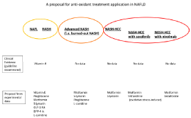
Dietary supplements, role and mechanism: Tables 3 and 4 and Figure 2

Figure 2: Showed [19]. Chemoprevention, role and mechanism: Tables 5 and 6.
Dietary supplement
Mechanism of action
Prevent hepatocarcinogenesis ,should include improvement in liver fibrosis
Dietary antioxidants coenzyme Q12, vitamin C and E, selenium
vE: A general cytotoxic ROS scavenger erases oxidative stress.
+ (short period studies).
Silymarin
is said to be an antioxidative agentRestored nicotinamide adenine
dinucleotide (NAD+) levels, and played a protective role against NAFLD.
Ameliorated liver fibrosis in NASHPotential HCC chemoprevention.
Supporting clinical evidence is lackingRegular coffee (>2 cups/day)
Reduced hepatocellular injury measured by IL6, ALT, AST,& GGT.
Meta analysis (RR 0.71)
case-control studies, (RR .53).
Tea intake , lessGreen tea
Reduction of DNA damage biomarkers
Undetermined
liquorice root
(HR 0.39)
Higher vitamin D, levels
(RR 0.51).
Patients with NASH have a deficiency of vitamin E and D. Vitamin D deficiency probably plays a role in hepatocarcinogenesis.
Table 3: Adapted from [3,12,17,18].
Dietary supplement
Mechanism of action
Prevent hepatocarcinogenesis
Unsaturated fat *PUFAs
It inhibit HCC growth through inhibition of COX2 and GSK-3b-mediated b-catenin degradation.
- (HR 0.71) * (HR 0.64)
white meat (chicken, turkey,
and fish)(HR 0.52)
red meat (beef and pork) (HR 1.74)Excessive dietary iron and/or genetic polymorphisms
Induce oxidative DNA damage and inflammation
increase HCC risk independently or alongside other aetiologies
BCAA
Reduces liver fibrosis
(RR 0.45)
L-carnitine
Controls the oxidative balance to aid the mitochondrial function, a fat-burning supplement.
Long-chain fatty acids taken up by mitochondria as complexes with L-carnitineanti-cancer agent
Table 4: Adapted from [12,19].
So, lack of a general consensus due to paucity of data makes us still do not know if it is Possible to Stop or Cease the NASH to Turn into HCC and increase its incidance. However Exercise, specially vigorous (RR of 0.56), Metformin (Reduced incidence 50% in diabetics) and Statins (In NAFLD without cirrhosis HR 0.29) may be helpful. Obeticholic acid, Elafibranor, Selonsertib and Cenicriviroc, the new drugs for NAFLD/NASH treatment we are waiting for, may carry more hope to decrease HCC (Table 5 and 6).
Dietary supplement
Mechanism of action
Prevent hepatocarcinogenesis
Unsaturated fat *PUFAs
It inhibit HCC growth through inhibition of COX2 and GSK-3b-mediated b-catenin degradation.
- (HR 0.71) * (HR 0.64)
white meat (chicken, turkey,
and fish)(HR 0.52)
red meat (beef and pork) (HR 1.74)Excessive dietary iron and/or genetic polymorphisms
Induce oxidative DNA damage and inflammation
increase HCC risk independently or alongside other aetiologies
BCAA
Reduces liver fibrosis
(RR 0.45)
L-carnitine
Controls the oxidative balance to aid the mitochondrial function, a fat-burning supplement.
Long-chain fatty acids taken up by mitochondria as complexes with L-carnitineanti-cancer agent
Table 5: Adapted from [3,6,12,17,19].
Therapy
Mechanism of action
histological features of NASH
Hepatocarcinogenesis
NB
NSAIDs,
including aspirinA pooled analysis of 10 cohorts suggested a protective effect of aspirin use
Phytochemicals: Silymarin- carotenoids -curcumin
Activate cytoprotective mechanisms such as the Keap1/Nrf2 pathway
Potential HCC chemoprevention.
Supporting clinical evidence is lacking.
Molecular targeted
Therapy
ACE& ARBsInhibit AGII mediated
NF-kB activation which promotes fibrogenic
myofibroblast survivalInhibit NASH fibrosis and HCC
Obeticholic acid,
*Elafibranor,
Selonsertib CenicrivirocFXR agonist
*A dual PPARa/d agonistImproved with all
Long-term benefits, safety, & its antihepatocarc- inogenesis need further evaluation
Probiotic, Prebiotic,
or synbiotic, or antibiotic treatmentAmeliorate IR- limit oxidative& inflammatory
liver damage - Reduce body fat& improve glucose metabolismImproved NASH activity index
Transplantation
Table 6: Adapted from [12,15].
References
- Abd Elkhalek Hamed, Medhat Elsahar, Nadia M Elwan, Sarah El-Nakeep, Mervat Naguib, Hanan Hamed Soliman, et al. Managing diabetes and liver disease association. Arab Journal of Gastroenterology. 2018; 19: 166–179.
- allestri S, Zona S, Targher G, Romagnoli D, Baldelli E, Nascimbeni F, et al. Nonalcoholic fatty liver disease is associated with an almost twofold increased risk of incident type 2 diabetes and metabolic syndrome. evidence from a systematic review and meta-analysis. J Gastroenterol Hepatol. 2016; 31: 936–944.
- Ahmet Uygun. Is That Possible to Stop or Cease the NASH to Turn into HCC?. J Gastrointest Canc. 2017; 48: 250–255.
- Wainwright P, Byrne CD. Bidirectional Relationships and Disconnects between NAFLD and Features of the Metabolic Syndrome. Int. J. Mol. Sci. 2016; 17: 367.
- Moore SW, Davidson A, Hadley GP, Kruger M, Poole J, Stones D, et al. Malignant liver tumors in South African children: a national audit. World J Surg 2008; 32: 1389–1395.
- Lung-Yi Mak, Vania Cruz-Ramón, Paulina Chinchilla-López, Harrys A Torres, Noelle K LoConte, John P Rice, et al. Global Epidemiology, Prevention, and Management of Hepatocellular Carcinoma. ASCO EDUCATIONAL BOOK. 2018.
- Jinjuvadia R, Patel S, Liangpunsakul S. The association between metabolic syndrome and hepatocellular carcinoma: systemic review and meta-analysis. J Clin Gastroenterol. 2014; 48: 172-177.
- Huikuan Chu, Brandon Williams, Bernd Schnabl. Gut microbiota, fatty liver disease, and hepatocellular carcinoma: Liver Research. 2018; 2: 43-51.
- Paradis V, Zalinski S, Chelbi E, Guedj N, Degos F, Vilgrain V, et al. Hepatocellular carcinomas in patients with metabolic syndrome often develop without significant liver fibrosis: a pathological analysis. Hepatology. 2009; 49: 851-859.
- Masao Omata, Ann-Lii Cheng, Norihiro Kokudo, Masatoshi Kudo, Jeong Min Lee, Jidong Jia, et al. Asia–Pacific clinical practice guidelines on the management of hepatocellular carcinoma: a 2017 update. Hepatol Int. 2017; 11: 317-370.
- Valter Donadon, Massimiliano Balbi, Pietro Casarin, Alessandro Vario, Alfredo Alberti. Association between hepatocellular carcinoma and type 2 diabetes mellitus in Italy: Potential role of insulin: World J Gastroenterol. 2008; 14: 5695–5700.
- Naoto Fujiwara, Scott L Friedman, Nicolas Goossens, Yujin Hoshida. Risk factors and prevention of hepatocellular carcinoma in the era of precision medicine: Journal of Hepatology. 2018 vol. 68- 526–549.
- Vilar-Gomez E, Chalasani N. Non-invasive assessment of nonalcoholic fatty liver disease: clinical prediction rules and blood-based biomarkers. J Hepatol. 2018; 68: 305-315.
- White DL, Kanwal F, El-Serag HB. Association between nonalcoholic fatty liver disease and risk for hepatocellular cancer, based on systematic review. Clin Gastroenterol Hepatol. 2012; 10: 1342–1359.
- Kerstin Schütte, Fathi Balbisi, Peter Malfertheiner. Prevention of Hepatocellular Carcinoma: Gastrointest Tumors. 2016; 3: 37-43.
- Hosaka T, Suzuki F, Kobayashi M, Seko Y, Kawamura Y, Sezaki H, et al. Long-term entecavir treatment reduces hepatocellular carcinoma incidence in patients with hepatitis B virus infection. Hepatology. 2013; 58: 98-107.
- Cholankeril G, Patel R, Khurana S, Satapathy SK. Hepatocellular carcinoma in non-alcoholic steatohepatitis. World J Hepatol. 2017; 9: 533-543.
- Said A, Ghufran A. Epidemic of non-alcoholic fatty liver disease and hepatocellular carcinoma. World J Clin Oncol. 2017; 8: 429-436.
- Uchida D, Takaki A, Adachi T, Okada H. Beneficial and Paradoxical Roles of Anti-Oxidative Nutritional Support for Non-Alcoholic Fatty Liver Disease: Nutrients. 2018; 10: 977.