Case Report
J Endocr Disord. 2020; 6(3): 1042.
Malignant Pheochromcytoma Presenting as a Huge Abdominal Mass in the Third Trimester of Pregnancy, a Case Report
Moslemi MK¹*, Nazer LSH¹ and Ghoddoosi M²
¹Department of Urology, Qom University of Medical Sciences, Iran
²Department of Pathology, Qom University of Medical Sciences, Iran
*Corresponding author: Mohammad Kazem Moslemi, Department of Urology, Kamkar Hospital, School of Medicine, Qom University of Medical Sciences, Qom, Iran
Received: December 01, 2020; Accepted: December 23, 2020; Published: December 30, 2020
Abstract
Introduction: Pheochromocytoma is a metabolically active tumor, which originates from the adrenal medulla chromaffin cells [1]. Five-year survival rate for the malignant pheochromocytoma is 40% [2]. Ten percent of all cases of pheochromocytoma are malignant which involves lymph nodes, liver, and lungs as the most common sites of metastasis [3]. Occurrence of pheochromocytoma during pregnancy is rare, and it is a high risk disease for the mother and fetus. Due to the variable clinical presentation, it needs a high rate of suspicion for its proper detection and treatment. Herein we present a huge right sided abdominal pheochromocytoma which was detected in the third trimester of pregnancy.
Case Presentation: The patient is a 27-year-old woman presented with a huge right sided pheochromocytoma that was found in the third trimester of pregnancy. Three months after an elective caesarian section, the tumor was eradicated completely and the patient’s condition was improved significantly in short term. Persistence of the hypertension and follow up imaging revealed recurrence of the tumor in the primary surgical bed that necessitated the reoperations and palliative chemotherapy 3 years after the primary surgery.
Discussion: Pheochromocytoma is a catecholamine-producing tumor that occurs in in one or both adrenal glands (85%) or involves the sympathetic ganglia (15%) [4]. It is one of the etiologies of hypertension with the prevalence of 0.1-0.6% [5]. The occurrence of pheochromcytoma in pregnancy is very rare with the rate of one in 54000 pregnancies [6,7] or frequency of 0.002% [8]. Malignant pheochromocytoma is radiologically and histologically similar to its benign counterparts [1].
Conclusion: In the case of appropriate follow up, 5 years survival is possible in operated pheochromocytoma even in the malignant and recurrent form of it. However, based on the literature data, this is the first reported case of postpartum malignant pheochromocytoma reported from the Iran.
Keywords: Hypertension; Pregnancy; Pheochromocytoma
Case Presentation
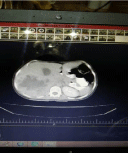
A previously well 27-year-old multiparous Afghanian refugee woman presented with a new onset hypertension and hyperglycemia during the third trimester of her pregnancy. The patient also complained of weight loss, anorexia and headache. She was referred to an obstetric center for management of her presumed preeclampsia but further investigations revealed nothing in favor of this disease. The patient remained hypertensive despite the hypertensive therapy. Considering huge right sided heterogenous mass, which was found in abdominopelvic ultrasonography (187*95*82 mm) and was expanding from liver to the pelvis, high suspicion of adrenal tumor was raised. Also high levels of catecholamine metabolites (metanephrine and Normetanephrine) were detected in the blood and 24 hour urine sample, which confirmed the probable diagnosis. An elective cesarean section was performed after a normal course of pregnancy and a healthy term baby was delivered. After several weeks the patient was admitted to the best equipped hospital of the city for resection of the tumor. Contrast enhanced abdominopelvic CT scan revealed a huge right sided retroperitoneal tumor extending from the sub hepatic area till the pelvic cavity (Figure 1).

    Figure 1: Subhepatic tumor with central necrosis is evident in the patient’s
abdominopelvic CT scan.
Admission blood pressure was 150/90 mmHg and the patient was on hypertensive medications including Diltiazem and Hydrochlorothiazid. The preoperative assessments such as EKG, CXR and routine lab studies showed no remarkable findings. After wide consultation with a multidisciplinary team consists of urologist, anesthesiologist, radiologist and cardiac surgeon the surgery was done on 4 May 2016. Despite the advantages of laparoscopic operation, we opted for an open approach due to the huge size of the tumor. We started the surgery through a midline incision, while entering the retroperitoneal space the mass was found extended downward beneath the liver. After dissecting the attachments to the surrounding tissues and organs, massive blood oozing was so hard to control that caused almost 7-8 liter of blood loss. The right adrenalectomy and tumor resection was successfully done but we had to leave the patient’s wound opened up and packed it with the use of 6 gauzes and 4 surgicels to limit the blood oozing from the posterior side of the liver. The patient was transferred to ICU after this 6 hour challenging surgery, 2 days later another laparotomy was done, the previous gauzes were taken out but due to the persistence of blood, oozing 36 surgicels were placed in the cavity. In the postoperation period fluctuation in the BP occurred with a maximum level of 200/150 mmHg. BP settled following administration of different oral antihypertensive drugs such as Labetalol, Trinitroglycerin, Hydrochlorothiazide, Diltiazem, Captopril, and Phenoxybenzmine. Also the Hemoglobine level which decreased to 8.5 g/dl was compensated by perfusion of several units of blood products such as packed RBCs and fresh frozen plasma. Eventually the patient was discharged on the seventh postoperative day with BP of 130/90 mmHg and prescription of 5 mg of Prazosin Tab, Bid. In the subsequent follow ups and after three years relapse of the tumor in the primary site with abdominal lymphadenopathies was happened which led to reoperation, but due to severe adhesions, tumor removal was impossible. Finally MIBG therapy was done and chemo-therapy was initiated with C is platine and Etoposide. The patient is still alive following her therapeutic schedule with close follow up.
Pathological Findings
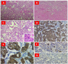
Gross examination of tumor showed large and lobulated mass, soft to firm in consistency with variegated cut surface, areas of hemorrhage necrosis and cystic change. Microscopic examination (Figure 2, A-C) showed relatively uniform tumor cells with abundant fine, granular cytoplasm, extensive necrosis, mitoses (up to 3/10 hpf) and focal periadrenal adipose tissue invasion. Immunohistochemical study showed the tumor cells are diffusely positive for Chromogranin and Synaptophysin and are negative for Inhibin. S100 immunostain highlighted presence of sustentacular cells around tumor nests (Figure 2, D-G). A final diagnosis of pheochromocytoma was made.

    Figure 2: Pheochromocytoma, microscopic and IHC findings.
A: Low power view of H&E stained section of tumor shows characteristic
nested (Zellballen) arrangement of tumor cells. B: Extensive necrosis of
tumor tissue (lower half). C: High power view of H&E stained section of
tumor shows tumor cells are relatively uniform with abundant fine, granular
red-purple cytoplasm. Mitosis present (inset). IHC study shows diffuse
cytoplasmic staining for Chromogranin. D: Synaptophysin. E: highlights
sustentacular cells. F&G: negative for Inhibin.
According to Pheochromocytoma of the Adrenal Gland Scaled Score (PASS), a total score of “6” was considered for this tumor. A PASS = or >4 indicates a potential for a biologically aggressive behavior.
Discussion
In the case of undiagnosed or untreated pheochromcytoma in pregnancy it has a fetal and maternal mortality of 40-50 % [9,10]. Invasion of adjacent structures or presence of multifocal disease can be one of determinants of malignancy of pheochromocytoma [1]. Our case has a large tumor with severe adhesions to adjacent structures such as liver and inferior vena cava. Due to huge size of tumor, Determination of successful tumor resection in our case was challenging. We did these preoperative, intraoperative and postoperative measurements for our case. Preoperative control of hemodynamic stability and cardiac arrhythmia, team working with the cardiac surgeon for increasing the chance of complete tumor removal and prevention of injuries to the neighborhood organs such as liver, and significant attention for the decreasing the rate of intraoperative blood loss and hemodynamic instability. Lastly close post-operative follow up surveillance was done appropriately. The potential nature of the tumor for infiltration, and its nature of high vascularity, makes the total removal of the tumor difficult and thus recurrence is common. We experienced significant intraoperative bleeding and multiple units of packed RBC and whole blood perfusion due to the highly vascular nature of the tumor. For the reduction of the risk of tumor recurrence adjuvant treatment such as chemotherapy, radiotherapy or MIBG therapy is recommended for the most of patients [11], but our case rejected it. One of determinants of metastatic pheochromocytoma is immunoreactivity for chromogranin A and synaptophysin [12], but our case did not undergo immunohistochemistry due to financial constraints.
Conclusion
Ten percent of pheochromocytoma is malignant, and recurrence of the tumor is one criteria of malignant pheochromocytoma. Follow up postoperative MIBG radionuclide imaging or abdominal ultrasonography is recommended for the detection of recurrent or metastatic tumors. In spite of huge size of primary tumor in our case and late initiation of follow up and chemotherapy, our case is still alive 4 years after diagnosing of malignant pheochromocytoma.
References
- Luis S, Song A, Zhou X, Kong X, Li WA, Wang Y, et al. Malignant pheochromocytoma with multiple vertebral metastases causing acute incomplete paralysis during pregnancy: Literature review with one case report. Medicine (Baltimore). 2017; 96: e8535.
- Lenders JW, Eisenhofer G, Manelli M, Pacak K. Pheochromcytoma. Lancet. 2005; 366: 665-675.
- Kaloostian PE, Zandik PL, Kim JE, Groves ML, Wolinsky J-P, Gokaslan ZL, et al. High incidence of morbidity following resection of metastaic pheochromocytoma in the spine. J Neurosurg Spine. 2014; 20: 726-733.
- Kaloostian PE, Zandik PL, Awad AJ, McCarthy E, Wolinsky J-P, Sciubba DM. En bloc resection of a pheochromocytoma metastatic to the spine for local tumor control and for treatment of chronic catecholamine release and related hypertension. J Neurosurg Spine. 2013; 18: 611-616.
- Lenders JW, Eisenhofer G, Manelli M, Pacak K. Pheochromocytoma. Lancet. 2005; 366: 665-675.
- Lenders JW. Pheochromocytoma and pregnancy: a deceptive connection. Eur J Endocrinol. 2012; 166: 143-150.
- Harrington JL, Farely DR, Van Heerden JA, Ramin KD. Adrenal tumours and pregnancy. World Journal of Surgery. 1999; 23: 182-186.
- Harper MA, Murnaghan GA, Kennedy L, Hadden DR, Atkinson AB. Pheochromocytoma in pregnancy. Five cases and a review of the literature. British Journal of Obstetrics and gynecology. 1989; 96: 594-606.
- Saathi V, Lila AR, Bangar TR, Menon PS, Shah NS. Pheochromocytoma and pregnancy: a rare but dangerous combination. Endocr Pract. 2010; 16: 300-309.
- Schenker JG, Chowers I. Phaeochromocytoma and pregnancy. Review of 89 cases. Obstetrical and Gynecological Survey. 1971; 26: 739-747.
- Dean RE. Pheochromocytoma and pregnancy. Obstetrics and Gynecology. 1958; 35-42.
- Adler JT, Meyer-Rochow GY, Chen H, Benn DE, Robinson BG, Sippel RS, et al. Pheochromocytoma: current approaches and further directions. Oncologist. 2008; 13: 779-793.
- Lau D, La Marca F, Camelo- Piragua S, Park P. Metastatic paraganglioma of the spine: case report and review of the literature. Clin Neurol Neurosurg. 2013; 115: 1571-1574.