Case Report
Austin ENT Open Access. 2018; 2(1): 1007.
Late Metastasis from Breast Cancer Mimicking Primary Chronic Osteomyelitis of Jaw: A Case Report
Hanc D¹*, Altun A² and Dinç S³
¹Department of Otolaryngology, Okmeydani Training and Research Hospital, Istanbul, Turkey
²Department of ENT clinics, Yunus Emre Hospital, Istanbul, Turkey
³Department of Radiology Department, Memorial Hospital, Istanbul, Turkey
*Corresponding author: Hanc D, Department of Otolaryngology, Okmeydani Training and Research Hospital, Istanbul, Turkey
Received: January 08, 2018; Accepted: February 12, 2018; Published: February 22, 2018
Abstract
Breast cancer is the most frequently diagnosed cancer in females. Even in early stage breast cancer, probability of local recurrence and metastasis in long period should be considered. We present a case CT and MRI findings were identified as an unusual presentation of late metastases of breast cancer. An 80-year-old female with previous history of breast cancer 16 years ago presented with vertigo and malnutrition. She had swelling, paresthesia on the right side of the cheek and difficulty in chewing. There wasn’t any sign of infection. Diffuse osteosclerosis without lysis and adjacent soft tissue swelling was seen on the right side of the mandible on CT scans. No fistula tract was identified. Cortical thickening, low signal intensity on T1 weighted images and high signal intensity on T2 weighted images of bone marrow was identified on MRI scan. Buccal, gingival and masetter muscle swelling were noted. These radiologic findings are more compatible with primary chronic osteomyelitis rather than breast cancer metastasis. Biopsy is needed for diagnosis.
The purpose of this report is to describe late metastases of breast cancer mimicking primary chronic osteomyelitis and its differential diagnosis in the mandible.
Keywords: Breast Carsinoma; Mandible; Metastasis; Osteomyelitis
Introduction
Metastatic tumors to the oral region are not common. Metastatic lesions may occur in the oral soft tissues, in the jawbones or in both osseous and soft tissue. Oral cavity metastases are considered rare and represent approximately 1% of all oral malignansies. Because of their rarity and atypical clinical and radiographic apperance, metastatic lesions are consider a diagnostic challenge. Because the most common jaw symptom is pain, these lesions could be misdiagnosed as pathologic entities with dental origin. Brest canser is one the most frequent canser in women worldwide. Brest canser is also the most frequent neoplasm that can metastasise to the head and neck region. In women the most common metastatic malignancies arise from primary cancers in from the breasts (42%), adrenals (8.5%), genital organs (7.5%) and thyroid glands (6%), but in men they arise from the lungs (22.3%), prostate (12%), kidneys(10.3%), bone (9.2%) and adrenals (9.2%). The lung cancer is the most common cancer that metastases to the oral soft tissues, whereas the breast cancer is the most common for metastatic tumors to the jawbones. The mandible is the most common location for metastases, with the molar area being the most frequently involved site in the jaw bones [1-4]. These sites are considered vulnerable to the deposition of neoplastic cells because of the presence of hematopoietic bone marrow, branching of the local blood vessels and slowing of blood flow [4].
Metastatic tumors of the oral cavity do not exhibit a pathognomonic radiographic appearance; therefore, radiographic examination is rarely considered diagnostically important. The diagnosis of a metastatic lesion in the oral region is challenging. Although most patients are previously diagnosed with primary neoplasms and treated, in one-third of the metastases the oral region presents the first clinical sign of the malignancy [5].
Patients with metastatic jaw disease demonstrate various clinical signs and symptoms that include commonly pain, swelling, paresthesia of the lip, loose or extruded teeth, halitosis, gum irritation, trismus, regional lymphadenopathy, mandibular nerve involvement and numb-chin syndrome, cortical expansion of the jawbones, ulceration, and exophytic growth [1,2,4]. Numbness or paresthesia of the lower lip and chin is considered an important sign of metastatic disease [5].
The clinical presentation simulates common pathologic conditions such as toothache, osteomyelitis, inflammatory hyperplasia, temporomandibular joint pain, trigeminal neuralgia, periodontal conditions, pyogenic, or giant cell granuloma, and accordingly, it may be difficult to diagnose such cases.
In the initial stages of the disease, the lesion may not produce a radiographic appearance. In an analysis of 390 cases of metastatic tumors of the jaw, Hirshberg et al. found that 5.4% of them did not show any important radiographic change [5].
The clinical appearance and course of primary chronic osteomyelitis makes diagnosis more difficult compared with cases of acute and secondary chronic osteomyelitis. A predisposing event, such an oral surgical procedure or an infectious tooth is missing. Clear signs of infection, such as pus or fistula formation are lacking.
The purpose of this report is to describe late metastases of breast cancer mimicking primary chronic osteomyelitis and its differential diagnosis.
Case Presentation
An 80-year-old female with a history of breast cancer 16 years admitted to our ENT clinic with complaints of difficulty in feeding, fatigue and subtle pain in the area of right mandibular molar parts. She had numbness for 3 years and swelling for 1 year on the right side of the cheek. In the medical anamnesis, it was discovered that the patient was operated (modified radical mastectomy with axillary lymph node dissection) for breast carcinoma of the right breast 16 years ago. After operation the patient did not take radiotherapy and chemotherapy treatment. The patient did not take bisphosthatase treatment. She had visited her physician 5 years periodically for annual examination. The patient’s medical history included diabetes and hypertension, and there was no history of tobacco or alcohol use. Clinical examination was difficult because of severe trismus. An intra–oral examination diffuse swelling, hard in palpation was observed on the right half of the mandibula. Movement of the jaw was restructed. There was severe pain over the right half of the face and paresthesia of the lower lip and chin. Regional lymph nodes were not palpable.
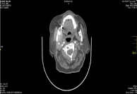
Axial and serial cross-sectional 1 mm-thick Cone Beam Computed Tomography (CBCT) showed small radiolucent areas in close proximity to the third molar (Figure 1) that were not diagnostic of metastases.

    Figure 1:  Adenocarcinoma: This tumor forms irregüler-shaped and a
cribriform glands with cytologically malignant cells exhibiting hiperchormatic
nuclei in a fibroblastic stroma.
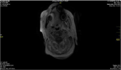
Magnetic Resonance Imaging (MRI) provided the most ccurate view of a cronic osteomielitis lesion in the right mandibula (Figure 2).

    Figure 2:  Magnetic Resonance Imaging (MRI) provided the most ccurate
view of a cronic osteomielitis lesion in the right mandibula.
Incisional biopsies were taken under local anesthesia from the mandibula and sent to the pathology laboratory.
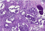
Five micron-thick, formalin-fixed, paraffin-embedded tissue sections stained with hematoxylin and eosin. The neoplastic cells contained abundant eosinophilic cytoplasm and large, pleomorphic, darkly stained nuclei (Figure 3). Several mitoses were observed, including atypical forms, as well as minimal lymphoplasmacytoid inflammatory infiltration of the stroma. The diagnosis was consistent with metastatic carcinoma of breast origin. Slides from the primary breast lesion were not available for comparison with the metastatic focus, because we did not find it.

    Figure 3:  Several mitoses were observed, including atypical forms, as well as
minimal lymphoplasmacytoid inflammatory infiltration of the stroma.
The patient was referred to the oncologist for further management, but the patient did not request any treatment. The patien died of her disease 6 month later.
Discussion
Metastasis is a consequence of complex biological cascade that begins with detachment of tumor cells from the primary tumor, spreading into the tissues, invading the lymphovascular structures followed by their survival in the circulation [5,6]. The microvasculature of the target organ provides room for the lodgment of metastatic tumor cells, from where they can extravasate, invade and proliferate within this target tissue. Angiogenesis is mandatory for the tumor cell load beyond 2-3 mm for adequate supply of oxygen and nutrients [7].
The diagnosis of metastasis to the oral cavity is a significant challenge to the clinician because of the lack of pathognomonic signs and symptoms.
Breast cancer primarily metastasizes to the regional lymph nodes, bone, lungs, pleura, and liver. Bone is the most common site of recurrence of the breast cancer which can be lytic, sclerotic or mixed. In the present case, the mandibular lesion was of sclerotic type. Sclerotic bone metastases are more common in patients with prostate, bladder, medulloblastoma or bronchial carcinoid tumors.
Late metastasis is usually defined for lesions when they appear more than 5 years after the treatment of a primary malignant tumor. Late recurrence of some of the malignant tumors such as renal cell carcinoma, breast cancer and malignant melanoma has been well documented in the literature. Late recurrences are common in Estrogen Receptor (ER) positive breast cancers. More than half of the recurrences of ER positive breast cancers occur 5 years or more after the diagnosis and treatment of the primary tumor, and some cases may recur even more than 20 years after the surgery [8]. In contrast to ER positive breast cancers, ER negative breast cancer recurrences are more common in the first two years of treatment and these are rare 5 years after the treatment [9].
The present case was an ER negative breast cancer and her mandibular lesion was diagnosed as late metastasis, occuring 16 years after the treatment of the primary tumor.
Oral region metastatic tumors are rare, comprising 1-3% of all malignant oral neoplasms. Malignant tumors of the breast, lung and kidney are the frequent primary sources. Metastatic tumors may occur in the oral soft tissues, in the jaw bones or both. Early detection of metastasis is important especially in oral metastasis where the prognosis is usually poor; most patient die within 1 year of diagnosis of oral metastasis, while the 4- year survival rate is estimated to be 10% [10,11]. The survival periods of patients with lung cancers were longer than those of patients with non- pulmonary tumors. Most patients with oral metastases have already developed generalized metastases by the time of diagnosis; however, in many cases, a solitary mandibular metastasis can be the initial manifestation of the primary tumor.
Mandibula is the frequently involved location for metastases. Clinical findings of mandibular metastasis may mimic reactive or benign lesions or sometimes simple odontogenic infections. Bony swelling with tenderness, pain, ulcer, hemorrhage, tooth mobility, trismus, paresthesia and pathological fracture can be seen as clinical findings [12].
According to some metaanalyses, the gender distribution of oral metastases is either predominantly male [10] approximately 2:1, respectively) or almost equal (approximately 1.1:1, respectively). [13,14]. In the Western literature, the most commonly reported primery site is the lung for males and the brest for females [10,13]. In this study we reported female patient.
Metastasis to the jaw bones occurs due to hematogenous route of the spread of malignant tumor and this requires the presence of hematopoietically active bone marrow well connected with sinusoidal vascular spaces at the site of deposition of malignant cells [15,16]. The posterior mandible and focal osteoporotic bone marrow defects in the edentulous mandible have been shown to be the hematopoietically active sites that may attract metastatic tumor cells [17,18]. Vascular changes associated with inflammatory process have been thought to be responsible for oral metastasis. Studies have shown that chronic trauma to the oral tissues favors metastatic spread of malignant tumors to the oral cavity [19]. The jaws do not contain a lymphatic system and experts think that metastasis occurs via the blood stream. The Batson’s vertebral venous plexus has been mentioned as a possible
Route of metastasis to the maxillofacial area, explaining why the lungs are not involved in some cases, as in this patient. In another study, it was found that in 55 cases, tooth extraction preceded the discovery of the metastasis [20]. Thus, the role of trauma to the oral mucosa, especially from ill-fitting denture, sharp tooth or restorations, poor oral hygiene and tooth extraction trauma, in the causation of oral metastasis needs further investigation.
Symptoms of oral/orofarengial metastases are variable and are not pathognomonic. When an oral lesion is found, even considering a benign condition, a biopsy should be mandatory, especially in patients with a known malignant disease.
Paresthesia of the lower lip and chin is the major symptom suggestive of metastatic disease. It is described in the literature as mental nerve neuropathy or Numb Chin Syndrome (NCS) [21,22]. The nerves associated with the NCS are the inferior alveolar nerve and its terminal branch, the mental nerve, which are branches of the third (mandibular) division of the trigeminal nerve. Because this nerve has no motor fibers, NCS is a sensory neuropathy. In addition to the chin and lip paresthesia, numbness of the teeth and mucosa may occur. The main cause of NCS includes perineural spread of metastatic disease or compression of nerve tissues by a tumor. Neoplasms that are most commonly associated with NCS are lymphomas and metastatic carcinomas of the mandible [22,23]. Although NCS may be iatrogenic and is often caused by dental anesthesia or inferior alveolar nerve injury after improper placement of dental implants, it may also occur as the result of acute or chronic osteomyelitis, odontogenic or nonodontogenic tumors or cysts, trauma, and also systemic diseases such as sickle cell anemia, multiple sclerosis, diabetes mellitus, and human immunodeficiency virus infections [24-26]. Our patient did not report paresthesia as the chief complaint, but careful intra-oral and extra-oral examinations revealed altered sensation to the lip and chin. Therefore, the existence of NCS should always alert the dentist or the physician to investigate the presence of a primary or recurrent malignant neoplasm, especially in cases that involve a significant medical history.
Numb chin syndrome may be the first and only symptom of an undergoing malignancy. Therefore, it should be regarded as malignancy until proven otherwise, and it should never be dismissed as a trivial symptom. Evaluation of a numb chin in a patient with a known malignancy should include a thorough physical examination to take out causes, followed by panoramic radiography, computed tomography or magnetic resonance imaging, and whole-body bone scan for detailed examination [27].
Osteomyelitis of the mandibula is classified as acute osteomyelitis, secondary chronic osteomyelitis and primary chronic osteomyelitis. Acute and secondary chronic osteomyelitis of the mandibula is essentially the same disease, arbitrarily separated by one month from the beginning of symptoms.
Secondary chronic osteomyelitis of the jaw may be associated with trauma, surgical procedures and dental infections such as endodontic and periapical infections [28].
Primary chronic osteomyelitis has no acute phase and results from a chronic low-grade infection of unknown cause. Constitutional symptoms and leukocytosis are usually absent in primary form.
Osteosclerosis is the most striking radiologic pattern seen in primary chronic osteomyelitis which is usually encountered in adult cases. Osteosclerosis and osteolysis can be seen together and leading to a so called “mixed pattern” which is more frequent in younger patients. Bone expansion and “onion skin” periosteal reaction are some of the radiologic features associated with primary chronic osteomyelitis. Sequestration is not seen any case of primary osteomyelitis [29]. Sclerotic form of the primary chronic osteomyelitis may be confused with fibrous dysplasia or Paget diseas [30].
In our current case the sclerotic bone lesion was compatible with late metastasis from breast cancer and primary chronic osteomyelitis. Biopsy and histopathological examination was required because clinical and imaging findings were unspecific for definitive diagnosis. Histopathological examination revealed ER negative breast cancer metastasis.
Conclusion
Otolaryngologists and dentists should include mandibular metastasis in the differential diagnosis during their general physical examination for patients with atypical symptoms, particularly if there is history of previous breast cancer even many years after the initial diagnosis.
Consent
Written informed consent was obtained from the patient for publication of this case report and any accompanying images.
References
- Dib LL, Soares AL, Sandoval RL, Nannmark U. Breast metastasis around dental implants: a case report. Clin Implant Dent Relat Res. 2007; 9: 112-115.
- D’Silva NJ, Summerlin DJ, Cordell KG, Abdelsayed RA, Tomich CE, Hanks CT, et al. Metastatic tumors in the jaws: a retrospective study of 114 cases. J Am Dent Assoc. 2006; 137: 1667-1672.
- Friedrich RE, Abadi M. Distant metastases and malignant cellular neoplasms encountered in the oral and maxillofacial region: analysis of 92 patients treated at a single institution. Anticancer Res. 2010; 30: 1843-1848.
- Akinbami BO. Metastatic carcinoma of the jaws: a review of literature. Niger J Med. 2009; 18: 139-142.
- Hirshberg A, Leibovich P, Buchner A. Metastatic tumors to the jawbones: analysis of 390 cases. J Oral Pathol Med. 1994; 23: 337-341.
- Hirshberg A, Buchner A. Metastatic tumours to the oral region: An overview. Oral Oncol Eur J Cancer Br. 1995; 31: 355-360.
- Hanahan D, Weinberg RA. The hallmarks of cancer. Cell. 2000; 100: 57-70.
- Dunnwald LK, Rossing MA, Li CI. Hormone receptor status, tumor characteristics, and prognosis: a prospective cohort of breast cancer patients. Breast Cancer Res. 2007; 9: 6.
- Zhang XH, Giuliano M, Trivedi MV, Schiff R, Osborne CK. Metastasis dormancy in estrogen receptor-positive breast cancer. Clin Cancer Res. 2013; 19: 6389-6397.
- Hirshberg A, Shnaiderman-Shapiro A, Kaplan I, Berger R. Metastatic tumours to the oral cavity: pathogenesis and analysis of 673 cases. Oral Oncol. 2008; 44: 743-752.
- Kesting MR, Loeffelbein DJ, Holzle F, Wolff KD, Ebsen M. Male breast cancer metastasis presenting as submandibular swelling. Auris Nasus Larynx. 2006; 33: 483-485.
- Kumar G, Manjunatha B. Metastatic tumors to the jaws and oral cavity. J Oral Maxillofac Pathol. 2013; 17: 71-75.
- Van der Waal RI, Buter J, van der Waal I. Oral metastases: report of 24 cases. Br J Oral Maxillofac Surg. 2003; 41: 3-6.
- Lim SY, Kim SA, Ahn SG, Kim HK, Kim SG, Hwang HK, et al. Metastatic tumours to the jaws and oral soft tissues: a retrospective analysis of 41 Korean patients. Int J Oral Maxillofac Surg. 2006; 35: 412-415.
- Kricum ME. Red -Yellow marrow conversion, its effect on the location of some solitary bone lesions. Skeletal Radiol. 1985; 14: 10-13.
- Morgan JW, Adcock KA, Donhouse RE. Distribution of skeletal metastasis in prostatic and lung cancer: Mechanism of skeletal metastsasis. Urology. 1990; 36: 31-34.
- Hashimoto N, Kurihara K, Yamasaki H, Ohba S, Sakai H, Yoshida S, et al. Pathological characteristics of metastatic carcinoma in the human mandible. J. Oral Pathol. 1987; 16: 362-367.
- Standish SM, Shafer WG. Focal osteoporotic bone marrow defects of the jaws. J Oral Surg. 1962; 20: 123-128.
- Monkman GR, Orwoll G, Ivinis JC. Trauma and oncogenesis. Mayo Clin Proc. 1974; 49: 157-163.
- Hirshberg A, Leibovich P, Horowitz I, Buchner A. Metastatic tumors to post-extraction site. J Oral Maxillofac Surg. 1993; 51: 1334-1337.
- Ryba F, Rice S, Hutchison IL. Numb chin syndrome: an ominous clinical sign. Br Dent J. 2010; 208: 283-285.
- Lesnick JA, Zallen RD. Numb chin syndrome secondary to metastatic breast disease. J Colo Dent Assoc. 1999; 78: 11-14.
- Harris CP, Baringer JR. The numb chin in metastatic cancer. West J Med. 1991; 155: 528-531.
- Yoshioka I, Shiiba S, Tanaka T, Nishikawa T, Sakamoto E, Kito S, et al. The importance of clinical features and computed tomographic findings in numb chin syndrome: a report of two cases. J Am Dent Assoc. 2009; 140: 550-554.
- Bar-Ziv J, Slasky BS. CT imaging of mental nerve neuropathy: the numb chin syndrome. AJR Am J Roentgenol. 1997; 168: 371-376.
- Smith SF, Blackman G, Hopper C. Numb chin syndrome: a nonmetastatic neurological manifestation of malignancy. Oral Surg Oral Med Oral Pathol Oral Radiol Endod. 2008; 105: 53-56.
- Narendra H, Ray S. Numb chin syndrome as a manifestation of metastatic squamous cell carcinoma of esophagus. J Cancer Res Ther. 2009; 5: 49-51.
- Elerson Gaetti-Jardim Júnior, Francisco Isaak Nicolas Ciesielski, Ricélia Possagno, Alvimar Lima de Castro, Antonio Carlos Marqueti & Ellen Cristina Gaetti-Jardim. Chronic Osteomyelitis of the Maxilla and Mandible: Microbiological and Clinical Aspects. Int. J. Odontostomat. 2010; 4: 197-202.
- Baltensperger M, Grätz K, Bruder E, Lebeda R, Makek M, Eyrich G, et al. Is primary chronic osteomyelitis a uniform disease? Proposal of a classification based on a retrospective analysis of patients treated in the past 30 years. J Craniomaxillofac Surg. 2004; 32: 43.
- Curé JK, Vattoth S, Shah R. Radiopaque jaw lesions: an approach to the differential diagnosis. Radiographics. 2012; 32: 1909-1925.