Case Report
Austin ENT Open Access. 2020; 3(1): 1009.
Floor of Mouth Neurofibroma: A Rare Presentation
Stansfield J1*, Sharma S1, Mirza O2 and Jayaram S1
¹Fairfield General Hospital, Northern Care Alliance, Manchester, UK
²Memorial Sloane Kettering Hospital, New York City, USA
*Corresponding author: Joseph Stansfield, Ear, Nose and Throat (ENT) Surgical trainee, North West Deanery, Department of ENT, Fairfield General Hospital, Northern Care Alliance, Bury, UK
Received: October 30, 2020; Accepted: December 15, 2020; Published: December 22, 2020
Abstract
Solitary sporadic neurofibromas are uncommon benign tumours of the neural sheaths of the peripheral nervous system. Solitary sporadic neurofibromas of the oral cavity and floor of mouth are extremely rare. We present the case of a 36-year-old male who presented with a 3-month history of a soft non-tender floor of mouth mass. MRI demonstrated a cystic septated lesion filling the left sublingual space. The lesion was excised via an external approach. Histology and immune assay unexpectedly demonstrated a neurofibroma.
Keywords: Sporadic neurofibroma; Floor of mouth; Case report
Background
Neurofibromas are common benign slow-growing tumours of the peripheral nervous system; they are well-circumscribed masses and compromise, in varying amounts, of Schwann cells, perineural cells and endo-neural fibroblasts [1]. It is the most common benign tumour of the peripheral nervous system. Neurofibromas can be solitary or multiple and can form part of a generalized syndromes such as Neurofibromatosis-1 (NF1) (Von Recklinghausen Syndrome) which presents as multiple neuro-cutaneous lesions due to an autosomal dominant mutation [2]. Oral neurofibromas occur in around 4-7 % of patients with a generalized neurofibroma syndromes such as NF1 [3]. However, Solitary sporadic neurofibromas unrelated to a syndrome presenting in the floor of mouth and oral cavity are extremely rare with only a few documented cases. Clinical presentation is similar to other benign oral cavity lesions and differential diagnosis includes plunging Ranula, dermoid cyst, and thyroglossal cyst amongst others. We present the case of a 36-year-old male with a three-month history of a soft, non-tender, mass that had progressively increased in size.

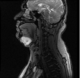
Figure 1: T2 weighted sagittal view MRI.

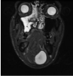
Figure 1: STIR weighted coronal view MRI.

Figure 3: T1 weighted coronal view MRI.
Case Presentation
A 36-year-old male presented to the outpatient clinic with a 3-month history of swelling in the submental region. It had gradually increased in size with occasional discomfort but no specific pain and the patient was otherwise asymptomatic. The patient had no significant past medical history and there was no family history to suggest a hereditary cause. On examination, there was a 5x5 cm mobile soft non-tender mass in the submental region. Although this lump could be felt on bimanual palpation, there was no obvious lesion visible in the floor of mouth. The patient had good dental health with no suspicion of an odontogenic cause for the swelling. The differential diagnosis of lipoma, dermoid tumour, thyroglossal cyst or a plunging ranula were considered. A contrast Magnetic Resonance Imaging (MRI) scan of the neck was organised in the first instance.
Investigations
The MRI scan demonstrated a well-defined septated cystic lesion filling the left side of the floor of mouth in the left sublingual space measuring 5x4x3.7 cm, descending inferiorly below the mandible compatible with a Ranula arising from the left sublingual space. T1 weighted imaging demonstrated low-intermediate intensity signal, whilst T2 weighted imaging demonstrated high intensity in keeping with other reported cases [3,4,9]. The left submandibular gland and right floor of mouth were unremarkable.
Treatment
The patient was listed and consented for an external approach excision of the mass with excision of the sublingual gland. At operation a large bilobed cystic swelling was identified causing significant bulging through the mylohyoid. No invasion of surrounding structures was noted. The cyst was resected en-bloc along with the sublingual gland. The specimen was sent for histological analysis.
Outcome and Follow-up
Histology reported the macroscopic appearance of a cyst. The lesion had two separate lobes measuring 42x40x17 mm and a second smaller lobe measuring 25x30x12 mm connected by a fibrous strand. Microscopy showed haphazard collagen deposition with spindle cells in a myxoid stroma. There was no evidence of atypia. The cells were strongly positive for S-100 and focally positive with NSE and NF. This immune profile suggested neurofibroma over differentials such as myxoid schwannoma. Overall, the pathology was suggestive of a benign nerve sheath tumour most likely in keeping with a neurofibroma. The patient made an uneventful postoperative recovery and no neurological deficit was noted. Patient was subsequently discharged.
Discussion
Oral neurofibromas are extremely rare and floor of mouth neurofibromas even more so. According to Broly et al., there have been 27 reported cases of neurofibroma in the oral cavity of which there have been 5 documented cases in the floor of mouth and submandibular region [3-7]. In these cases, the lesions had been suspected of arising from the terminal branches of the lingual nerve having been traced to the main trunk [3,5]. Despite the neural involvement neurological deficit is very rare in oral cavity solitary sporadic neurofibroma with only one case demonstrating any neurological deficit, (dystheasia in a patient with a tongue neurofibroma) [8]. Due to the lack of focal neurology, identifying the specific nerve involved if not identified intraoperatively can be challenging. Floor of mouth neurofibromas may be difficult to diagnose both clinically or radiologically as they may mimic both benign and malignant lesions in this region. An increase in size of the lesion may cause symptoms due to local compression. Ultrasound or MRI remain the best imaging modalities to both detect and demonstrate the extent of the neurofibroma in the oral cavity and floor of mouth region.
Evidence exists of malignant transformation of neurofibromas in patients with NF1 and NF2, and although there are fewer reports of this in solitary sporadic neurofibromas, this should still be taken into consideration10. Due to this potential risk, and the need to confirm a histological diagnosis, complete surgical excision of the lesion should be recommended. This can be performed both through an external or, if amenable, via a transoral approach [4]. Although the option of transoral approach was considered for this patient, we eventually decided to use an external incision because of lack of discrete lesion visible in the floor of mouth. The potential of long-term loss of neurological function post operatively should be taken into consideration when consenting the patient. In conclusion, sporadic solitary neurofibromas of the floor of the mouth are extremely rare. Due to the diagnostic challenges with both clinical examination and imaging, and the potential for malignant transformation if left untreated, surgical excision of the lesion should be undertaken.
Acknowledgement
Thanks are owed to all ENT clinical staff who were involved in this patients care.
References
- Srivathsa S. Solitary neurofibroma of the floor of the mouth. International Journal of Clinicopathological Correlation. 2017; 1: 20-22.
- Sakata A, Hirokawa Y, Kuwahara R, Hamada A, Kuroda M, Araki N, et al. Solitary oropharyngeal neurofibroma: MR appearance with pathologic correlation and review of the literature. Clin. Imaging. 2013; 37: 554-557.
- 3. Broly E, Lefevre B, Zachar D, Hafian H. Solitary neurofibroma of the floor of the mouth: rare localization at lingual nerve with intraoral excision. BMC Oral Health. 2019; 19: 197.
- Maruyama M, Fushiki H, Watanabe Y. Solitary neurofibroma of the floor of the mouth: a case report. Case Rep. Otolaryngol. 2011; 2011; 967896.
- Wang HM, Hsu YC, Lee KW, Chiang F-Y, Kuo W-R. Neurofibroma of the lingual nerve: a case report. Kaohsiung J Med Sci. 2006; 22: 461-464.
- Chao YT, Li WY, Chu PY. Postradiation Tumor in the Floor of Mouth. JAMA Otolaryngol. Head Neck Surg. 2015; 141: 663-664.
- Al-Omran MK, Al-Khamis ANK, Malik AK. Solitary neurofibroma of the floor of the mouth. NeuroSci Riyadh. 2006; 11: 53-55.
- Lykke E, Noergaard T, Rasmussen ER. Lingual neurofibroma causing dysaesthesia of the tongue. BMJ Case Rep. 2013; 2013: bcr2013010440.
- Pilavaki M, Chourmouzi D, Kiziridou A, Skordalaki A, Zarampoukas T, Drevelengas A. Imaging of peripheral nerve sheath tumors with pathologic correlation: pictorial review. Eur. J. Radiol. 2004; 52: 229-239.
- Kamra HT, Dantkale SS, Birla K, Sakinlawar PW, Bharia PH. Plexiform neurofibroma in the submandibular gland along with small diffuse neurofibroma in the floor of the mouth but without neurofibromatosis: a rare case report. Ecancermedicalscience. 2013; 7: 313.