Case Report
Austin ENT Open Access. 2024; 4(1): 1012.
Managing Magnetic Resonance Imaging Artifacts for Patients with Cochlear Implant
Arkoubi Z*; Benkhraba N; Bencheikh R; Benbouzid A; Oujilal A; Essakalli L
Department of ENT, Head and Neck Hospital of Rabat, Morocco
*Corresponding author: Arkoubi Z Department of ENT, Head and Neck Hospital of Rabat, Morocco. Email: zakaria.arkoubi@gmail.com
Received: February 21, 2024 Accepted: March 25, 2024 Published: March 29, 2024
Abstract
Nowadays, Magnetic Resonance Imaging (MRI) is an essential tool in the diagnostic process, mainly concerning the neurological pathology. Its innocuous nature, and the newest sequences make it more useful. However, some patients do not benefit from it advantages due to the metallic devices that they carry. For patients with cochlear implants, the manufacturer made it possible thanks to a great work on the magnetic field. The cochlear implant is actually no more an obstacle for MRI but the appearance of artifacts can in some cases make the interpretation impossible, and make surgery once again, the only way to reduce these artifacts. That’s what we are going to talk about in this article through a case of a 3 years old little girl who needed a surgery procedure to remove the magnetic device of the implant in a goal to reduce the artifacts.
Introduction
The cochlear implant is a revolutionary device that has a considerable positive impact on the psychological development of the youngest children. But till that day, we still need surgery for the implantation, which can in rare cases leads to neurological complications. This situation makes it a good reason to do an MRI for a patient with cochlear implant. This foreign body can be responsible for artifacts that can seriously complicate the interpretation. In this paper we are going to present a case of a 3-year-old girl with a cochlear implant who needed a surgical procedure, removing the magnetic device before the MRI test to reduce artifacts. Even if it’s a new generation device, artifacts can be an obstacle in a 1.5 Tesla MRI, especially for young children with cranial surface much lower than the adults.
Case
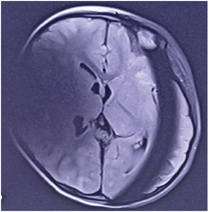
It’s about a 3 years old little girl that make no response to sonore stimulations observed by her parents, and also an absence of language. The patient had an electrophysiological test that diagnosed bilateral cophosis. The girl had an MRI in a goal to implant the right ear at first which diagnosed a Gusher malformation. The surgical procedure took more time than usual due to the leak of the LCS caused by the Gusher syndrome. Six months after the surgery, the patient was in the intensive care unit because of meningoencephalitis. During her stay in the intensive care unit, the patient didn’t wake up after the stop of the hypnotics. The MRI didn’t come up with conclusive results to explain her cerebral state, due to the importance of artifacts (Figure 1).

Figure 1: Artifacts in the first MRI.
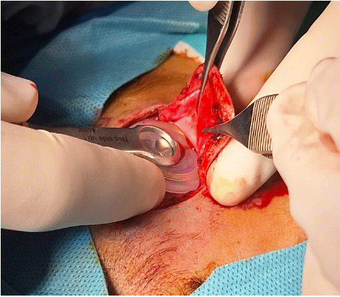
The MRI was done a second time but with no difference than the first one. A surgery was performed to remove the magnetic device and then replace it with a nonmagnetic one (Figure 2).

Figure 2: Replacement of the magnetic device with a specific tool.
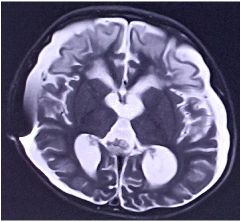
The artifacts were then less important than in the 2 first MRIs (Figure 3). A cerebral aplasia was diagnosed for this 3 years old little girl. The patient actually is out of the intensive care unit but still have psychosomatic deficiency.

Figure 3: MRI after removing the magnetic device.
Discussion
Actually, the profound bilateral deafness is not the only indication for the cochlear implantation. The latest researches in audiological sciences confirms that there are much more indications for cochlear implant than before, both in the pediatric and the adult population. Some of the patients with cochlear implant will certainly need an MRI in their lifetime. As an example, the patients with neurofibromatosis, with tumor in the cochlear nerve. Only the MRI can follow the evolution of their tumoral lesions. We will add that for those patients, the MRI in some cases can’t confirm if the nature of the tumor is benign or malignant [1]. From that last example, it’s clear that an implanted patient could need an MRI in its lifetime. To carry a cochlear implant nowadays doesn’t make it a contraindication for the MRI due to the work that has been done on the magnetic field. The diametral magnetic field can allow the magnetic device to stay stable during the realization of the MRI. This diametric magnetic field reduces also the artefacts, in fact Med EL presented the first device with diametral magnetic field in 2015 [2]. This discussion about the presence and the importance of the artifacts can open a debate on the size of the head related to the size of the implant. When an adult patient with cochlear implant is having an MRI, it’s certainly not as a 1- or 2-years old patient referring to the importance of the artifacts. But it must be proven by advanced studies. An artifact generated by a cochlear implant in a child head will maybe cover more volume than in adult patients, so should we deduce that this kind of surgical procedure is more frequent in the pediatric population? Unfortunately, there is not enough studies on the subject. However, it’s reported that the artifacts are present in 100% of patients during the MRI but the degree is not precised [2,3]. Our patient had 3 MRIs during her stay in the intensive care unit due to the artifacts. The third one, which was done after the surgical replacement of the magnetic device could let the radiologist get with a diagnostic, but that didn’t remove the artifact at a 100%. The cochlear implant is a foreign body, and every time there is a foreign body it generates artifact in contact with X rays or with a magnetic field. The question is that: should we make the surgical procedure systematically or should we give it a shot? For a patient like the one that we’ve talked about, it’s certainly not safe for her to multiply transports from the intensive care to the radiology unit, it could lead to more cerebral lesions. Making the surgical procedure at first could avoid 3 transports. But for a “healthy” patient, the MRI could be done at first, and should we let the surgery for cases when the artifacts don’t permit the interpretation of the images? Especially that this kind of surgery could be done also with local anesthesia [4]. Once again only more advanced studies could afford responses.
Conclusion
Till that day the artefacts are still here as long as there is a foreign body. The technology worked on the magnetic device of the cochlear implant and came up with the diametric magnetic field that reduced these artifacts and make it possible for a patient with a cochlear implant to have an MRI. The question about making the surgical procedure at first or let it once the artefacts couldn’t make an interpretation possible will need more advanced researches. Anyways, we can be grateful that today, patients with cochlear implant could have an MRI without any complication.
References
- Neurofibromatosis 1 french National Guidelines based on an extensive literature review since 1966 Christina Berggyist et al Orphanet Journal of rare Diseas. 2020; 15: 37.
- Young NM, Hoff SR, Ryan M. Impact of cochlear implant with Diametric Magnet on imaging Acess, safety and clinical care Nancy M. Young et Al Laryngoscope. 2021; 131: E952- E956.
- Specific considerations for determining safety with MRI use in cochlear implant patients Douglas D et Al Cochlear implant, An update. 2002; 45–48.
- Migirov L, Wolf M. Magnet removal and reinsertion in a cochlear implant recipient undergoing brain MRI. ORL J Otorhinolaryngol Rela spec. 2013; 75: 1-5.