Case Report
Austin ENT Open Access. 2024; 4(1): 1015.
Unilateral Nasal Obstruction Revealing a Pharyngeal Venous Malformation: A Case Report
Ayyad K1,2*; Dahan T1,2; El Mekkaoui M1,2; El Hafi Z1,2; Arkoubi Z1,2; Bencheikh R1,2; Benbouzid A1,2; Oujilal A1,2; Essakalli L1,2
1Department of Otorhinolaryngology and Neck-Face surgery Hospital of Specialities Rabat, Morocco
2Faculty of Medecine and Pharmacy of Rabat, Mohammed V University Rabat, Morocco
*Corresponding author: Ayyad K Department of Otorhinolaryngology and Neck-Face Surgery, Mohammed V University, Rabat, Morocco. Tel: 212678123018 Email: Ayyad.ka@gmail.com
Received: April 13, 2024 Accepted: May 07, 2024 Published: May 14, 2024
Abstract
Venous malformations are part of slow-flow vascular tissue anomalies. Cervicofacial localization is common, but pharyngeal involvement is rare. We report the case of a 17-year-old patient, who is being treated for Crohn’s disease and asthma, presenting to the emergency department with a significant painful naso and oropharyngeal mass evolving for 6 weeks. This mass caused unilateral nasal obstruction and difficulty swallowing. Nasofibroscopy revealed a violaceous budding process in the nasopharynx covered with venous lacis, pedunculated behind the right tubal orifice, extending towards the oropharynx, lining its right lateral wall, pushing the soft palate and uvula to the left, and reaching the posterior pillar of the right tonsil and the apparently healthy epiglottis. In addition to laboratory tests, a facial mass CT scan described a right-sided pharyngeal lesion.The patient underwent a complete surgical excision of the mass through a combined endoscopic endonasal and endobuccal approach. The final histological result favored a venous malformation with no signs of malignancy. The 3-month follow-up showed good local control. Pharyngeal venous malformations are rare and can be serious, potentially jeopardizing vital prognosis if large. T2-weighted MRI is key for diagnosis. Treatment involves sclerotherapy, considered the gold standard, with surgery retaining certain indications, either alone or in combination with sclerotherapy.
Keywords: Venous Malformation; Pharynx; Rare; Sclerotherapy; Surgery; Case Report
Introduction
Vascular anomalies are classified by the International Society for the Study of Vascular Anomalies into two major families: vascular tumors and vascular malformations. Venous malformations are part of slow-flow vascular tissue anomalies. Cervicofacial localization is common. They can be superficial or deep, potentially affecting vital prognosis.
Clinical Observation
A 17-year-old, followed for Crohn's disease with Immurel for a year and a half, asthmatic under crisis treatment only, presented to the emergency department with the appearance, over 6 weeks, of a significant painful naso- and oropharyngeal mass causing unilateral nasal obstruction and swallowing discomfort, associated with right hemicranial pain, without epistaxis, and evolving in a context of weight loss.
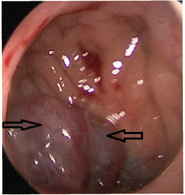
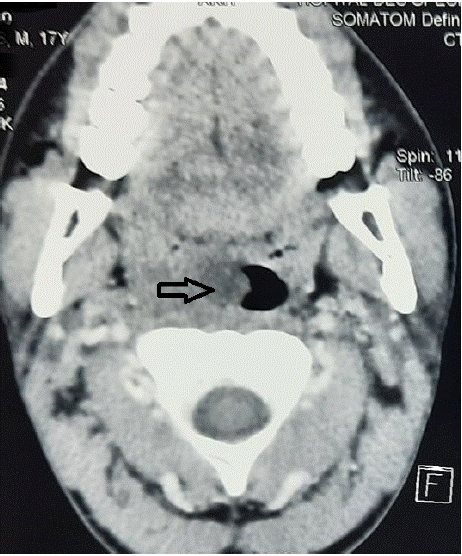
Nasofibroscopy revealed a violaceous budding process in the nasopharynx covered with venous lacis, pedunculated behind the right tubal orifice, extending towards the oropharynx, lining its right lateral wall, pushing the soft palate and uvula to the left, reaching the posterior pillar of the right tonsil and the apparently healthy epiglottis (Figure 1). Additionally, examinations of lymph node areas, neurological examination, and the rest of the ENT and general examinations were unremarkable. Blood tests, including complete blood count and coagulation profile, showed no abnormalities. Facial CT described a right-sided oropharyngeal lesion (Figure 2).

Figure 1: Endoscopic aspect of violaceous process in the nasopharynx.

Figure 2: Facial CT scan image showing the orophayngeal mass.
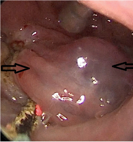
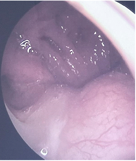
After initial embolization, the patient underwent complete excision of the mass through a combined endoscopic endonasal and endobuccal approach (Figure 3). The final histological result favored a venous malformation with no signs of malignancy. The 3-month local control was good (Figure 4).

Figure 3: Peroperative endoscopic aspect showing the excision of the process.

Figure 4: Postoperative endoscopic appearance at 3 months showing good local control.
Discussion
Venous malformations are the most common vascular malformations, often found in cervicofacial regions. They can affect various surfaces, including the oral cavity, tongue, lip, palate, larynx, or pharynx. According to Steiner et al [1], these malformations can occur in skin, subcutaneous tissue, muscles, or bones, with 4.6% of patients having pharyngeal or laryngeal involvement. They are usually sporadic, with only 1.2% being familial [2]. A mutation in the TIE-2 gene has been found in familial and 50% of sporadic cases [3]. These malformations are present from birth and may manifest after a period of quiescence, with their size increasing rapidly due to hormonal changes, trauma, increased venous pressure, revealing their presence [4]. Clinical presentation varies based on localization, ranging from a bluish, painful, depressible swelling that increases during Valsalva maneuvers in superficial forms to bleeding, dysphagia, dysphonia, orthopnea, sleep apnea, and upper airway obstruction in deep or pharyngo-laryngeal forms [5-7]. Clinical examination is complemented by nasofibroscopy for a comprehensive evaluation. Imaging, specifically MRI, assesses the extension, anatomical localization, and presence of satellite lesions. Venous malformations appear hyperintense on T2-weighted sequences. Contrast injection helps differentiate them from lymphatic malformations, which show progressive and heterogeneous enhancement [8]. Communication with a deep venous network should be investigated [9]. Blood tests are necessary due to potential coagulation disorders, often showing reduced fibrinogen and elevated D-dimers. In extensive forms, there is a risk of disseminated intravascular coagulation. Evolution can lead to painful inflammatory episodes, requiring NSAID treatment or more severe complications affecting vital prognosis due to mass effect on the upper airways. Therapeutic management depends on factors such as extension, depth, and risk of airway obstruction. Superficial lesions, especially mucosal ones, are treated with ND-YAG laser. For large lesions with superficial components, initial ND-YAG laser treatment may induce submucosal fibrosis, followed by sclerotherapy or surgery, typically 4 to 6 weeks after laser treatment [10]. Percutaneous sclerotherapy can also be used for small to medium-sized lesions, employing agents such as 95% ethanol, bleomycin, Trombovar, Aetoxisclerol, or Polidocanol to induce fibrosis and destruction of the venous malformation's endothelium. Inflammation, cutaneous or mucosal necrosis, muscle fibrosis, and compressive edema can complicate sclerotherapy [11]. According to several studies, belomycin can be used safely and effectively without tracheotomy via a transmucosal endoscopic approach [12]. Surgery is indicated for extensive/giant lesions, in conjunction with sclerotherapy for small lesions (<3 cm), allowing complete excision, for thrombosed lesions where sclerotherapy is ineffective, or after the failure of sclerotherapy. Surgery, when indicated, should be performed away from any inflammatory episodes and ideally starting from the age of 2. Partial excision of venous malformations may require monitoring combined with sclerotherapy or repeated surgeries for voluminous lesions [13].
Conclusion
Pharyngeal venous malformations are rare and can be serious, potentially jeopardizing vital prognosis if large. T2-weighted MRI is key for diagnosis. Treatment involves sclerotherapy, considered the gold standard, with surgery retaining certain indications, either alone or in combination with sclerotherapy.
References
- Frederica Steiner, Kiarash Taghavi, Trevor FitzJohn, Swee T. Tan. Stratification and characteristics of common venous malformation by anatomical location. JPRAS Open. 207; 13: 29-40.
- Boon LM, Mulliken JB, Enjolras O, Vikkula M. Glomuvenous malformation (glomangioma) and venous malformation: distinct clinicopathologic and genetic entities. Arch Dermatol. 2004; 140: 971-976.
- Limaye N, Wouters V, Uebelhoer M, Tuominen M, Wirkkala R, Mulliken JB, et al. Somatic mutations in angiopoietin receptor gene TEK cause solitary and multiple sporadic venous malformations. Nat Genet. 2008; 41: 118-124.
- Pappas Jr DC, Persky MS, Berenstein A. Evaluation and treatment of head and neck venous vascular malformations. Ear Nose Throat J. 1998; 77: 914-916, 918-22.
- Clarke C, Lee EI, Edmonds J Jr. Vascular anomalies and airway concerns. Semin Plast Surg. 2014; 28: 104–10.
- Durr ML, Meyer AK, Kezirian EJ, Mamlouk MD, Frieden IJ, Rosbe KW. Sleep-disordered breathing in pediatric head and neck vascular malformations. Laryngoscope. 2017; 127: 2159–64.
- Teresa MO, Alexander RE, Lando T, Grant NN, Perkins JA, Blitzer A, et al. Segmental hemangiomas of the upper airway Laryngoscope. 2009; 119: 2242–7.
- Barbier C, Martin A, Papagiannaki C, Cottier J-P, Lorette G, Herbreteau D. Malformations veineuses superficielles ou «angiomes veineux» Superficial venous malformations. Presse Med. 2010; 39: 471–81.
- Chowdhury FH, Haque MR, Kawsar KA, Sarker MH, Momtazul Haque AFM. Surgical management of scalp arterio-venous malformation and scalp venous malformation: an experience of eleven cases. Indian J Plast Surg. 2013; 46: 98–107.
- Tristan Klosterman, Teresa MO. The Management of Vascular Malformations of the Airway Natural History, Investigations, Medical, Surgical and Radiological Management. Otolaryngol Clin N Am. 2018; 51: 213–223.
- Baud AV, Breton P, Guibaud L, Freidel M. Treatment of low-pressure vascular malformations by injection of Ethibloc. Study of 19 cases and analysis of complications. Rev Stomatol Chir Maxillofac. 2000; 101: 181–8.
- Oomen KP, Paramasivam S, Waner M, Niimi Y, Fifi JT, Berenstein A, et al. Endoscopic transmucosal direct puncture sclerotherapy for management of airway vascular malformations. Laryngoscope. 2016; 126: 205–11.
- Ethunandan M, Mellor TK. Haemangiomas and vascular malformations of the maxillofacial region: a review. Br J Oral Maxillofac Surg. 2006; 44: 263–72.