Case Report
Austin ENT Open Access. 2024; 4(2): 1016.
Cervical Vagus Nerve Schwannoma: A Case Report
Benyahia Z1,2*; EL Hafi Z1,2; Bencheikh R1,2; Benbouzid A1,2; Oujilal A1,2; Essakalli L1,2
1Department of ENT, Hospital of Specialties, Morocco
2University Mohammed V Rabat, Morocco
*Corresponding author: Zainab Benyahia Department of ENT, hospital of specialties, university Mohamed V Morocco. Email: zainab.benyahia1992@gmail.com
Received: April 13, 2024 Accepted: May 07, 2024 Published: May 14, 2024
Abstract
Cervical schwannomas are benign tumors that develop exclusively from the Schwann cells of peripheral nerves. They are rare, particularly when they affect the cervical vagus nerve. This article illustrates, through a clinical case, the distinctive imaging features and histological peculiarities of these tumors, underlining the importance of surgical treatment. We report the case of a 75-year-old man presenting with an isolated left parapharyngeal mass that appeared three months ago. Cervical imaging (CT and MRI) revealed a vascularized mass associated with a paraganglioma of the carotid glomus. Extracapsular surgical excision was performed by cervicotomy, uncovering a tumor arising from the left cervical vagus nerve, identified as a schwannoma on histological analysis. The post-operative period was without complications. When faced with an isolated parapharyngeal mass, it is crucial to consider vagus nerve schwannoma as a possible diagnosis. Preoperative imaging studies (CT and MRI) are essential to suggest this diagnosis. The therapeutic approach is based on surgical intervention, necessary both to confirm the histological diagnosis and to prevent recurrence, thanks to complete extracapsular excision.
Introduction
Schwannoma is a benign mesenchymal tumor that originates exclusively in the Schwann cells that form the sheath around the nerve fibers of the peripheral nervous system. 25% of schwannomas are found in the cervical region, typically associated with the vagus nerve (X) [1]. These tumors are notable for their slow development and often delayed diagnosis due to their less obvious clinical manifestation. The preferred management is surgical intervention, although the decision to proceed with surgery is carefully weighed against the potential risks of functional sequelae for the patient. In the context of a recent case of cervical vagus nerve schwannoma, this article aims to provide, through an in-depth review of the literature, an overview of the clinical features, radiological diagnostic methods, and therapeutic approaches to this uncommon anatomo-clinical pathology.
Patient and Observation
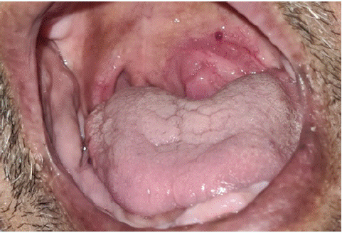
The patient was 75 years old, hypertensive and on treatment. For 3 months, he had presented with high dysphagia without dysphonia or dyspnea, evolving in a context of preservation of his general condition. Clinical examination revealed a left laterocervical curvature with an oropharyngeal bulge pushing back the homolateral tonsil (Figure 1).

Figure 1: Clinical appearance of the left laterocervical curvature and oropharyngeal bulge.
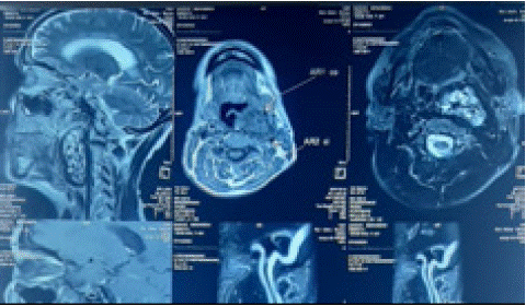
Cervical CT scan revealed a tumoral process of the palatine tonsil and the left parapharyngeal space (Figure 2).

Figure 2: Cervical CT scan showing the tumoral process of the palatine tonsil and left parapharyngeal space.
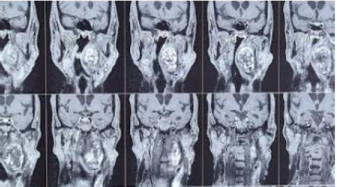
Cervico-facial MRI revealed a left latero-cervical lesional process, opposite the carotid bifurcation responsible for the reduction of the oropharyngeal lumen. Measuring 63x52x31mm, it was associated with a paraganglioma of the left carotid glomus (Figure 3).

Figure 3: Cervicofacial MRI revealing a left latero-cervical lesional process opposite the carotid bifurcation.
He underwent complete removal of the tumor, with a double team of ENT and vascular surgeons (Figure 4). The definitive pathology came back in favor of a schwannoma. The postoperative course was straight forward.
Discussion
The first description of a cervical schwannoma was made by Ritter in 1899. These tumors occur in the head and neck in 25-45% of cases [1,2]. However, they are more prevalent in the intracranial region, particularly on the vestibular nerve [3], while their presence in the cervical region is less common. These tumors can develop within the parapharyngeal space, affecting the last four cranial nerves as well as the cervical sympathetic nerve [4,5,6].
Vagus nerve schwannomas occur across all ages, with a more marked incidence in younger adults. The male/female ratio is generally balanced according to most studies [1,2], although some observations have reported a slight female predominance [7]. Clinically, these tumors often present as an isolated laterocervical or parapharyngeal mass, progressively enlarging and generally asymptomatic [2,4]. Depending on their size, they may compress the pharynx, causing discomfort or odynophagia [2,8]. A vagus nerve schwannoma may be suspected in the presence of a cervical mass accompanied by dysphonia and vocal cord paralysis on the same side, although this sign is rare at diagnosis due to nerve compression [1,4].
Imaging plays a central role in the diagnosis and treatment of these patients [1,4,6]. While ultrasound offers only limited specificity, Computed Tomography (CT) and Magnetic Resonance Imaging (MRI) provide precise details on the size and parapharyngeal location of the tumor, its relationship to surrounding vascular structures, including the internal and external carotids [6,9]. CT is particularly useful for excluding non-vascularized adenopathies, congenital cysts and paragangliomas, which are characterized by strong contrast enhancement as early as the arterial phase [1,2,10]. On MRI images, schwannomas present with variable intensity in T1 sequence and become hyperintense in T2, with significant enhancement after gadolinium injection [2,7]. Diagnostic confirmation is based on histopathological examination, revealing a proliferation of Schwann cells with a characteristic structure organized in Antoni A and B zones. S100 protein is frequently expressed, as shown by immunohistochemistry [1,11].
The preferred therapeutic strategy for schwannomas is surgery, which allows both diagnosis and resection of the tumor. Thanks to their well-encapsulated nature, total excision is often possible, with preservation of the nerve of origin in most cases [7,12]. In our case, a total extracapsular resection of the schwannoma was performed, with preservation of the vagus nerve. The most frequent post-operative complication is recurrent paralysis, especially in cases of X-nerve injury. The use of intraoperative neuromonitoring can be advantageous for surgery on cervical nerve tumors, offering the possibility of partial resection in cases of preoperatively diagnosed vagus nerve schwannoma.
Small, slow-growing cervical schwannomas, particularly those arising from the vagus nerve, greater hypoglossus or a branch of the cervical plexus, should prompt a reassessment of the indication for surgery [1].
The prognosis for patients with schwannomas is generally excellent. Nerve complications and local recurrence are rare, the latter probably due to incomplete excision [1,13].
Conclusion
In summary, cervical vagus nerve schwannoma is a benign but rare pathological entity that requires special attention due to its clinical and surgical implications. The case presented illustrates the importance of a rigorous diagnostic approach, highlighting the crucial role of advanced medical imaging, namely Computed Tomography (CT) and Magnetic Resonance Imaging (MRI), in the preoperative identification of these tumors. Extracapsular surgical excision has proved to be an effective strategy for confirming the histological diagnosis and avoiding recurrence. This case reinforces the notion that, although rare, vagus nerve schwannomas should be considered in the face of masses. Successful treatment relies on complete excision, which not only confirms the diagnosis but also ensures the absence of recurrence, thus contributing to a favorable prognosis for the patient. This experience highlights the importance of multidisciplinary collaboration and meticulous surgical planning to optimize results and minimize post-operative risks.
Author Statements
Informed Consent
A written informed consent, dated and signed, was obtained from the patient.
Conflict of Interest
The authors declare no conflict of interest.
Ethics Approval Obtained
This study was approved by: Mohammed V university in Rabat; morocco.
Authors' Contributions
All authors have contributed to the management of the patient and have read and approved the final version of the manuscript.
Acknowledgements
To all authors who contributed to the realization of this work.
References
- Nao EE, Dassonville O, Bozec A, Sudaka A, Marcy PY, Vincent N, et al. Cervical sympathetic chain schwannoma. Eur Ann Otorhinolaryngol Head Neck Dis. 2012; 129: 51–3.
- Chiofalo MG, Longo F, Marone U, Franco R, Petrillo A, Pezzullo L. Cervical vagal schwannoma. A case report. Acta Otorhinolaryngol Ital. 2009; 29: 33–5.
- Malone JP, Lee WJ, Levin RJ. Clinical characteristics and treatment outcome for nonvestibular schwannomas of the head and neck. Am J Otolaryngol. 2005; 26: 108–12.
- Furukawa M, Furukawa MK, Katoh K, Tsukuda M. Differentiation between schwannoma of the vagus nerve and schwannoma of the cervical sympathetic chain by imaging diagnosis. Laryngoscope. 1996; 106: 1548–52.
- Tomita T, Ozawa H, Sakamoto K, Ogawa K, Kameyama K, Fujii M, et al. Diagnosis and management of cervical sympathetic chain schwannoma: a review of nine cases. Acta Otolaryngol. 2009; 129: 324–9.
- Anil G, Tan TY. Imaging characteristics of schwannoma of the cervical sympathetic chain: a review of 12 cases. AJNR Am J Neuroradiol. 2010; 31: 1408–12.
- Kim SH, Kim NH, Kim KR, Lee JH, Choi HS. Schwannoma in head and neck: preoperative imaging study and intracapsular enucleation for functional nerve preservation. Yonsei Med J. 2010; 51: 938–42.
- Colreavy MP, Lacy PD, Hughes J, Bouchier-Hayes D, Brennan P, O’Dwyer AJ, et al. Head and neck schwannomas - a 10-year review. J Laryngol Otol. 2000; 114: 119–24.
- Saito DM, Glastonbury CM, El-Sayed I, Eisele DW. Parapharyngeal space schwannomas. Preoperative imaging determination of the nerve of origin. Arch Otolaryngol Head Neck Surg. 2007; 133: 662–7.
- Casserly P, Kiely P, Fenton JE. Cervical sympathetic chain schwannoma masquerading as a carotid body tumor with a postoperative complication of first-bite syndrome. Eur Arch Otorhinolaryngol. 2009; 266: 1659–62.
- Enzinger FM, Weiss SW. Benign tumors of peripheral nerves. In: Enzinger FM, Weiss SW, editors. Soft tissue tumors. 3rd ed. Saint Louis: Mosby. 1995; 821–872.
- Fujino K, Shinohara K, Aoki M, Hashimoto K, Omori K. Intracapsular enucleation of vagus nerve originated tumours for preservation of neural function. Otolaryngol Head Neck Surg. 2000; 123: 334–6.
- Gilmer-Hill HS, Kline DG. Neurogenic tumours of the cervical vagus nerve: report of four cases and review of the literature. Neurosurgery. 2000; 46: 1498–503.